Outreach and Education 
Video: How Enzymes Work
Every second inside every living cell, thousands of chemical reactions are taking place. These reactions constitute the essential tasks of life such as metabolism, protein synthesis, cell renewal and growth.
Explore the PDB-101 Browser to learn more about enzymes.Meeting Reports
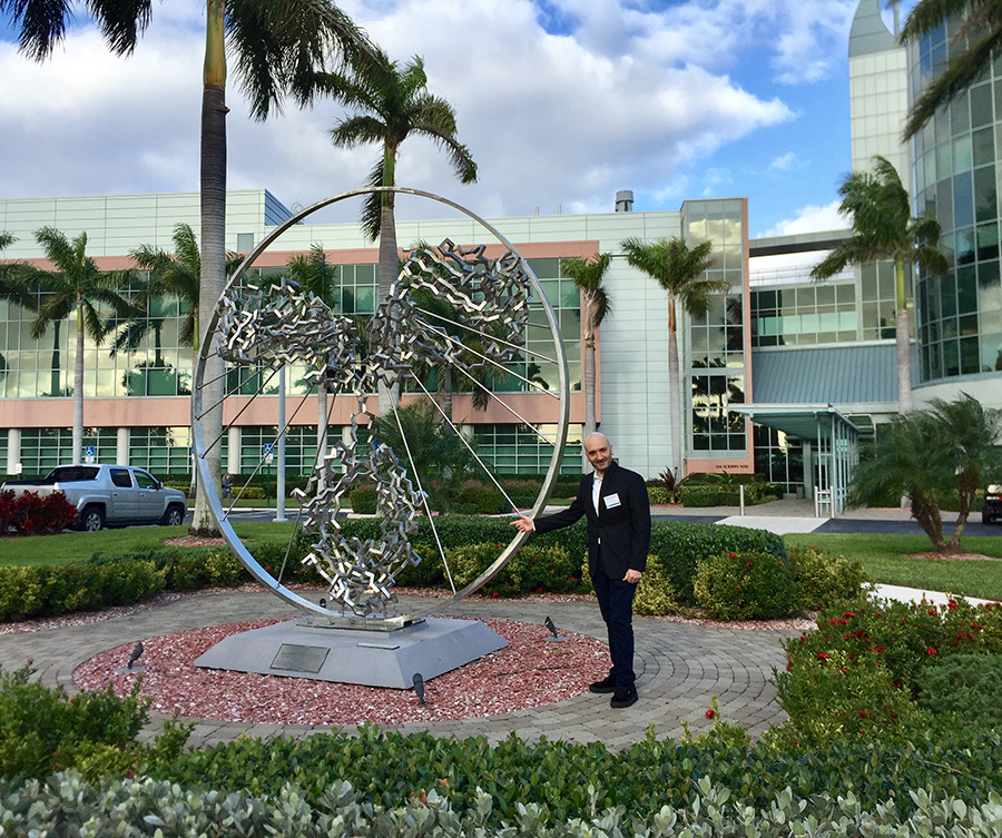
Luigi with the Angel of the West Julian Voss-Andreae at Scripps. Voss-Andreae also created the Synergy sculpture outside of the RCSB PDB offices at Rutgers.
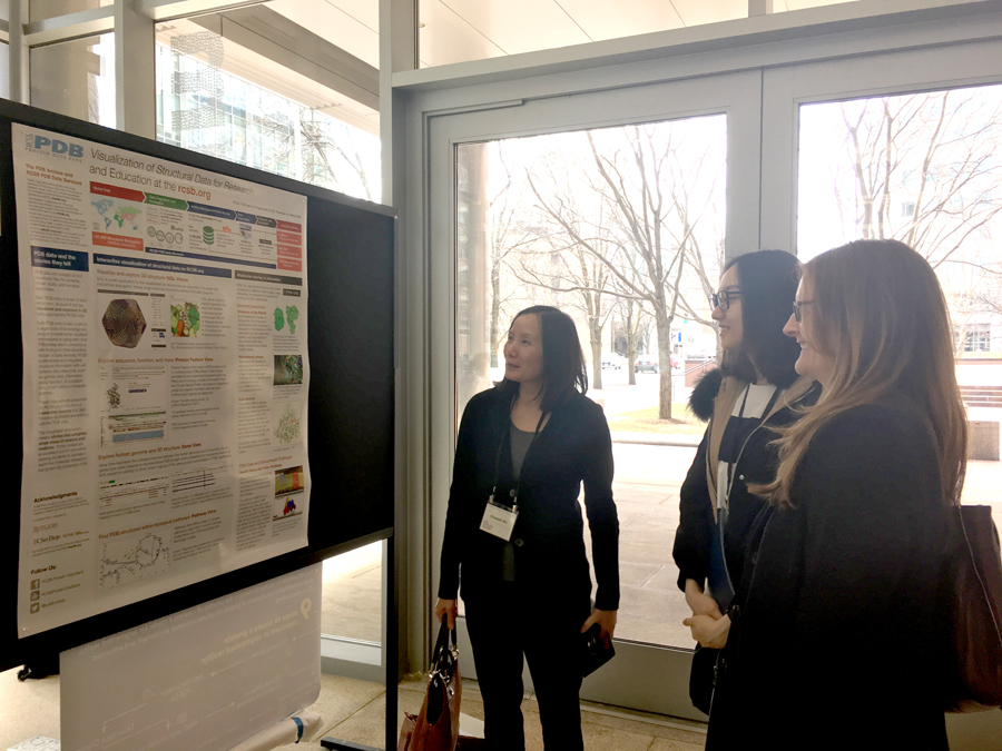
RCSB PDB Poster at VIZBI
Biocurator Luigi Di Costanzo described Biocurating the Molecules of Life for the Protein Data Bank at the AAAS Florida Biomedical Career Symposium organized by The Scripps Research Institute and the Max Planck Florida Institute in January. The symposium brought together from across the country to share their career experiences to mentor graduate students and postdoctoral fellows on post-Ph.D. career possibilities. Luigi had similarly described his biocuration work with Science Careers ("The rewards of working as a data wrangler" doi: 10.1126/science.caredit.aaq0481).
Biocuration Developer Chenghua Shao gave an overview of XFEL deposition and data architecture support in PDB at the 2018 BioXFEL conference and EuXFEL workshop in New Orleans in February. Serial femtosecond crystallography (SFX) and free electron X-ray lasers (XFEL) are revolutionizing the methods of X-ray crystallography. wwPDB is working with field-experts to develop data extensions to collect of XFEL-specific metadata and other XFEL-related improvements.
During February’s Biophysical Society Meeting (San Francisco), several interesting posters presented by Helen Berman and Brinda Vallat were on display, including Sustaining a living digital data resource that enables breakthroughs in scientific research and biomedical education; Interactive Exploration of Non-Covalent Interactions with the NGL Viewer; Towards a Common General Purpose Molecular Query Language; and A Data Dictionary and Prototype Deposition System for Archiving Integrative/Hybrid Models.
At the AAAS Annual Meeting (Austin, TX; February), Christine Zardecki described how RCSB PDB is Sustaining A Living Digital Data Resource That Enables Breakthroughs in Scientific Research and Biomedical Education during the poster session.
The 9th International Meeting on Visualizing Biological Data (VIZBI) was held this year March 28–30 at the Broad Institute of MIT and Harvard (Cambridge MA). Maria Voigt presented a poster on the Visualization of Structural Data for Research and Education at rcsb.org. An image depicting the alpha-amylase enzyme was part of the Art and Biology evening.
Art, Biology and Visualization
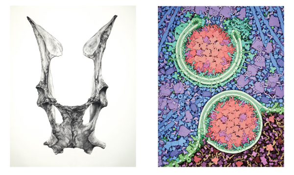
Left: Tara Shukla, Pelvic Bone, 2016. Charcoal on paper, 30 x 22 in. Courtesy of the artist. Photo by Justin Hayworth. Right: David Goodsell, Selective Autophagy 1,000,000X, 2016. Watercolor and ink on paper, 14 x 11 in. Courtesy of the artist; from the Molecule of the Month feature on Aminopeptidase 1 and Autophagy
Making Life Visible: Art, Biology, and Visualization explores the processes of visualization and description in art and biology by featuring work by 16 contemporary artists and scientists, as well as historical material from the 16th to 19th centuries. The exhibit will be on display at Faulconer Gallery at Grinnell College in Grinnell, Iowa from February 2 - June 10, 2018.
The show features works from Molecule of the Month creator David Goodsell, including paintings of Autophagy and Zika.
In the past, biologists and artists had similar training in observation and drawing. Though the fields have diverged, individual practitioners on both sides continue to draw inspiration from one another, finding new ideas in the process of creating images. The exhibition asks: what do artists and biologists see, and how do their ways of seeing challenge and stimulate one another? Exhibition subjects range from molecules and cells, to organisms and ecosystems, and the artists/scientists work in labs, studios, museums, and in the field.
The show is curated by Professor Jackie Brown, Biology Department and Dr. Lesley Wright, director, Faulconer Gallery and funded in part by a grant from the National Science Foundation (DEB-1457741).
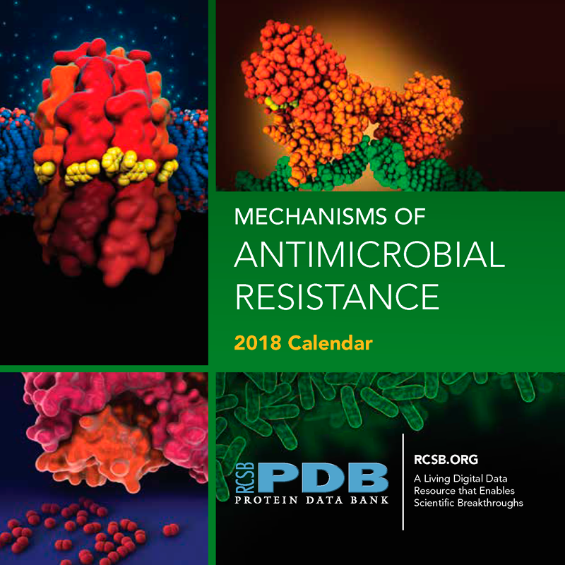
2018 Health Focus: Antimicrobial Resistance
Antibiotics have saved countless lives, but pathogens are quickly finding ways to survive antibiotic treatment. Antibiotic-resistant bacteria are predicted to become the leading cause of death worldwide, with an expected death rate of 10 million people annually by 2050. They take many approaches: pumping antibiotics out of their cells, altering the molecular machinery that the antibiotics target, and attacking the antibiotics directly. Atomic structures publicly available in the PDB are revealing the details of drug resistance and providing new ways to combat it.
RCSB PDB invites high school students across the USA to study the molecular mechanisms behind the resistance, create a short video, and submit to our annual challenge. To learn more, visit PDB-101.
The deadline for submissions is May 23. Award winners will be announced on June 12, 2018.
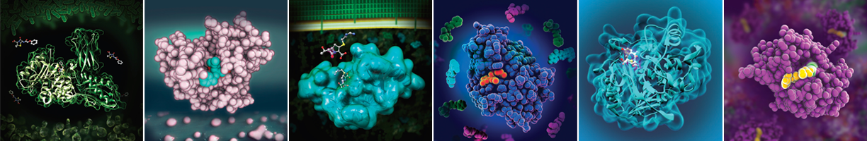
PDB-101 offers many other materials focused on Antimicrobial Resistance to help students in the 2018 Video Challenge, including this 2018 calendar.
This calendar was created by the RCSB PDB and Jenna Abyad (Drew University) and Priscilla Salcedo (California State University, Northridge) as part of a summer internship with RCSB PDB as described in our Newsletter.
Enter to win a printed copy by sending your address via the Contact Us button, or download in PDF or PowerPoint formats.