Data Exploration Services
RCSB.org Statistics
| Month | Unique Visitors | Visits | Bandwidth |
|---|---|---|---|
| July 2020 | 707,006 | 2,553,617 | 10566.80 GB |
| August 2020 | 682,088 | 2,162,671 | 4080.59 GB |
| September 2020 | 686,187 | 2,285,846 | 2679.69 GB |
Explore Sequence-Structure Relationships with PFV
Protein Feature View visually maps a PDB chain (or "instance") to corresponding sequences from UniProtKB and annotations from external resources in different "tracks" to enable explorations of structural and biological features.
This tool integrates
- Secondary structure, angle/distance outliers, protein-ligand binding sites or disulfide bridges (from PDB data)
- Structural domains (from CATH and SCOPe)
- Biochemical and biomedical features (from UniProtKB)

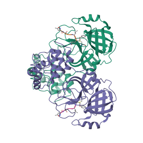
Protein Feature View for 3sn6. Zoom in with the mouse or two fingers for a granular view that includes amino acids; click to highlight specific areas; hold and click to move left/right.
The Macromolecules section on a Structure Summary Page provides a truncated Protein Feature View. Selecting the Sequence tab launches the expanded view for the structure.
Protein Feature View also maps a UniProt sequence to all corresponding structures in the PDB. Access this feature from the Macromolecules section on a Structure Summary Page or enter the UniProt ID in the URL rcsb.org/uniprot/. Visit the Help Documentation for complete details.
Integrating PDB Data with NIH Common Fund Data Resources
RCSB PDB has integrated PDB structural information with three NIH Common Fund Data Resources. Ultimately, this integration is expected to enable investigation of novel biological questions, and promote a more complete understanding of human health and disease.
- PHAROS is part of the Integrating the Druggable Genome (IDG) project and presents integrated data for 3 of the most important drug target protein families: GPCRs, ion channels and kinases. Some of the data types it covers include: disease, phenotypic and ontologies such as the Drug Target Ontology.
- GTEx is a public resource for the study of tissue-specific gene expression and regulation.
- IMPC covers data related to mouse phenotypes, as part of an international effort by 19 research institutions to identify the function of every protein-coding gene in the mouse genome.
Data are accessible from individual Structure Summary pages and Advanced Search.


Searching PDB entries that have a mapping to these NIH Common Fund data resources is possible in the RCSB.org Advanced Search User Interface by selecting Attribute: Linked External Resources and UniProt-mapped Resource.
This work was supported by an NIH Common Fund Data Use Award intended for Enhancing Utility and Usage of Common Fund Data Sets.
Search by Chemical Formula or Descriptor

Use the Advanced Search>Chemical Search option to find chemical components (standard and modified amino acids/nucleotides, and ligands) by Formula (exact match or portion) or SMILES or InChi descriptor (graph-relaxed, graph-relaxed-stereo, graph-strict, fingerprint-similarity). A description of this functionality is available.
Advanced Search Query Builder can be used combine these Chemical Searches with other types of searches using Boolean operators (AND/OR/NOT):
- Attribute searching: specific fields or full-text search across all fields, such as ID, deposition information, entry features, and experimental information
- Sequence searching: based on a FASTA sequence (with E-value or % Identity cutoffs);
- Sequence Motif searching: find short sequence patterns in PDB structure FASTA sequences using Simple, PROSITE, or RegEx syntax
- Structure Similarity searching: based on an existing Chain or Assembly of a PDB structure
The Attribute search can be used to find Chemical Components based on Name, Synonyms, Chemical Component Type, InChiKey, Molecular Weight, Atom Count, eternal database IDs, and binding affinity.
Advanced Search results can be returned as PDB structures, polymer entities, assemblies, or non-polymer entities that correspond to the search performed.
Small molecules in the PDB are described in the wwPDB Chemical Component Dictionary.
Retiring Legacy Services
Recent improvements to RCSB.org include revised and updated tools for searching and exploring PDB data. As a result, select RCSB PDB Services are being replaced or retired.
Updated: MyPDB Service

MyPDB accounts allow users to save search queries, rerun saved search queries, and receive email notifications when new structures matching saved queries are released.
To utilize the new and improved MyPDB features, users should generate a new MyPDB account using third-party authentication (Google, Facebook, or ORCID).
Legacy MyPDB users are encouraged to generate new MyPDB accounts. Legacy email notifications have been discontinued.
Retiring: Legacy RCSB PDB APIs
Recently-introduced Search and Data APIs offer comprehensive functionality and high performance. As a result, legacy RCSB PDB APIs (REST search and fetch) will be discontinued in November 2020.
Documentation is available to help users learn the concepts and syntax behind the new services. Use this documentation to display all of the details of a specific API request, and then run the search request and to see the response.
Updated: Molecular Graphics
Structure Summary Pages and Search Results have been refreshed with new images created using Mol*, the new 3D visualization tool. Any links to legacy images were retired. Users should update URLs to new images (e.g., http://cdn.rcsb.org/images/structures/lu/6lu7/6lu7_assembly-1.jpeg) before that date.