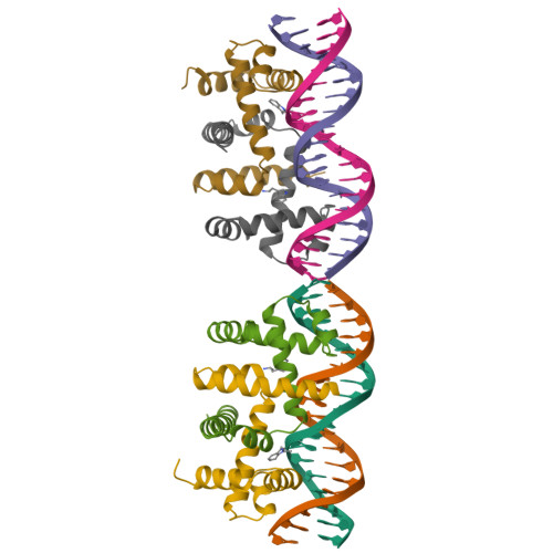
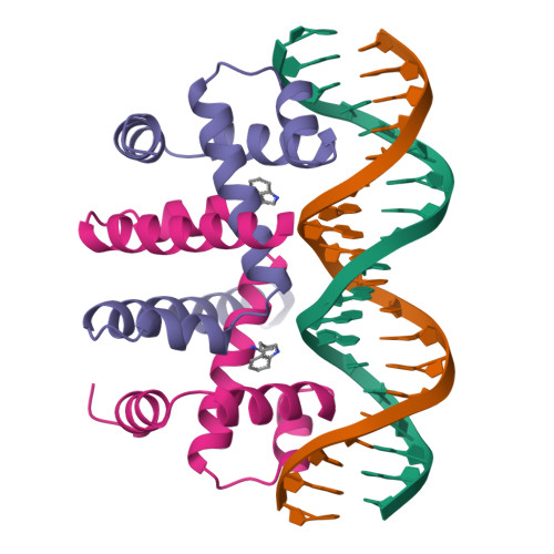
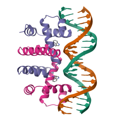
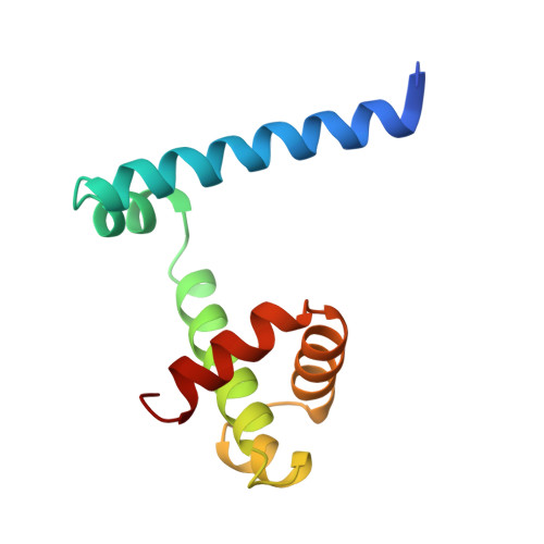
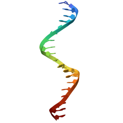
RCSB PDB - 1TRO: CRYSTAL STRUCTURE OF TRP REPRESSOR OPERATOR COMPLEX AT ATOMIC RESOLUTION

 1TRO

CRYSTAL STRUCTURE OF TRP REPRESSOR OPERATOR COMPLEX AT ATOMIC RESOLUTION


Experimental Data Snapshot

  • Method: X-RAY DIFFRACTION
  • Resolution: 1.90 Å
  • R-Value Observed: 0.167 

wwPDB Validation   3D Report Full Report


This is version 1.4 of the entry. See complete history


Literature

Crystal structure of trp repressor/operator complex at atomic resolution.

Otwinowski, Z.Schevitz, R.W.Zhang, R.G.Lawson, C.L.Joachimiak, A.Marmorstein, R.Q.Luisi, B.F.Sigler, P.B.

(1988) Nature 335: 321-329

  • DOI: https://doi.org/10.1038/335321a0
  • Primary Citation of Related Structures:  
    1TRO

  • PubMed Abstract: 

    The crystal structure of the trp repressor/operator complex shows an extensive contact surface, including 24 direct and 6 solvent-mediated hydrogen bonds to the phosphate groups of the DNA. There are no direct hydrogen bonds or non-polar contacts to the bases that can explain the repressor's specificity for the operator sequence. Rather, the sequence seems to be recognized indirectly through its effects on the geometry of the phosphate backbone, which in turn permits the formation of a stable interface. Water-mediated polar contacts to the bases also appear to contribute part of the specificity.


  • Organizational Affiliation

    Department of Biochemistry and Molecular Biology, University of Chicago, Illinois 60637.


Macromolecules

Find similar proteins by:  (by identity cutoff)  |  3D Structure
Entity ID: 2
MoleculeChains Sequence LengthOrganismDetailsImage
PROTEIN (TRP REPRESSOR)E [auth A],
F [auth C],
G [auth E],
H [auth G]
108Escherichia coli str. K-12 substr. W3110Mutation(s): 0 
UniProt
Find proteins for P0A881 (Escherichia coli (strain K12))
Explore P0A881 
Go to UniProtKB:  P0A881
Entity Groups  
Sequence Clusters30% Identity50% Identity70% Identity90% Identity95% Identity100% Identity
UniProt GroupP0A881
Sequence Annotations
Expand
  • Reference Sequence
Experimental Data & Validation

Experimental Data

  • Method: X-RAY DIFFRACTION
  • Resolution: 1.90 Å
  • R-Value Observed: 0.167 
  • Space Group: P 1 21 1
Unit Cell:
Length ( Å )Angle ( ˚ )
a = 43.65α = 90
b = 72.43β = 94.96
c = 107.39γ = 90
Software Package:
Software NamePurpose
PROLSQrefinement

Structure Validation

View Full Validation Report


Entry History 

Deposition Data

Revision History  (Full details and data files)

  • Version 1.0: 1994-01-31
    Type: Initial release
  • Version 1.1: 2008-05-22
    Changes: Version format compliance
  • Version 1.2: 2011-07-13
    Changes: Source and taxonomy, Version format compliance
  • Version 1.3: 2020-05-13
    Changes: Data collection, Database references, Structure summary
  • Version 1.4: 2024-02-14
    Changes: Data collection, Database references, Derived calculations