2BOP

CRYSTAL STRUCTURE AT 1.7 ANGSTROMS OF THE BOVINE PAPILLOMAVIRUS-1 E2 DNA-BINDING DOMAIN BOUND TO ITS DNA TARGET


Experimental Data Snapshot

  • Method: X-RAY DIFFRACTION
  • Resolution: 1.70 Å
  • R-Value Observed: 0.201 

wwPDB Validation   3D Report Full Report


This is version 1.4 of the entry. See complete history


Literature

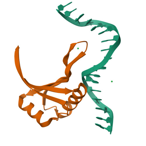
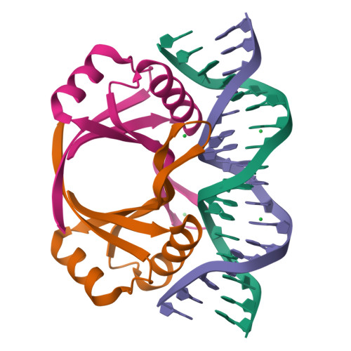
Crystal structure at 1.7 A of the bovine papillomavirus-1 E2 DNA-binding domain bound to its DNA target.

Hegde, R.S.Grossman, S.R.Laimins, L.A.Sigler, P.B.

(1992) Nature 359: 505-512

  • DOI: https://doi.org/10.1038/359505a0
  • Primary Citation of Related Structures:  
    2BOP

  • PubMed Abstract: 

    The dominant transcriptional regulator of the papillomaviruses, E2, binds to its specific DNA target through a previously unobserved dimeric antiparallel beta-barrel. The DNA is severely but smoothly bent over the barrel by the interaction of successive major grooves with a pair of symmetrically disposed alpha-helices. The specific interface is an 'interwoven' network of interactions where the identifying base pairs of the target contact more than one amino-acid side chain and the discriminating amino acids interact with more than one base pair.


  • Organizational Affiliation

    Howard Hughes Medical Institute, Yale University, New Haven, Connecticut 06510.


Macromolecules

Find similar proteins by:  (by identity cutoff)  |  3D Structure
Entity ID: 2
MoleculeChains Sequence LengthOrganismDetailsImage
PROTEIN (E2)B [auth A]85Deltapapillomavirus 4Mutation(s): 0 
UniProt
Find proteins for P03122 (Bovine papillomavirus type 1)
Explore P03122 
Go to UniProtKB:  P03122
Entity Groups  
Sequence Clusters30% Identity50% Identity70% Identity90% Identity95% Identity100% Identity
UniProt GroupP03122
Sequence Annotations
Expand
  • Reference Sequence

Find similar nucleic acids by:  Sequence   |   3D Structure  

Entity ID: 1
MoleculeChains LengthOrganismImage
DNA (5'-D(*CP*CP*GP*AP*CP*CP*GP*AP*CP*GP*TP*CP*GP*GP*TP*CP*G )-3')A [auth B]17N/A
Sequence Annotations
Expand
  • Reference Sequence
Small Molecules
Ligands 1 Unique
IDChains Name / Formula / InChI Key2D Diagram3D Interactions
YB
Query on YB

Download Ideal Coordinates CCD File 
C [auth B],
D [auth A]
YTTERBIUM (III) ION
Yb
AWSFICBXMUKWSK-UHFFFAOYSA-N
Experimental Data & Validation

Experimental Data

  • Method: X-RAY DIFFRACTION
  • Resolution: 1.70 Å
  • R-Value Observed: 0.201 
  • Space Group: R 3 2
Unit Cell:
Length ( Å )Angle ( ˚ )
a = 64.6α = 61.8
b = 64.6β = 61.8
c = 64.6γ = 61.8
Software Package:
Software NamePurpose
NUCLSQrefinement
PROFFTrefinement

Structure Validation

View Full Validation Report


Entry History 

Deposition Data

Revision History  (Full details and data files)

  • Version 1.0: 1994-01-31
    Type: Initial release
  • Version 1.1: 2008-05-22
    Changes: Version format compliance
  • Version 1.2: 2011-07-13
    Changes: Version format compliance
  • Version 1.3: 2019-11-20
    Changes: Advisory, Derived calculations
  • Version 1.4: 2024-02-14
    Changes: Data collection, Database references, Derived calculations