3CPA

X-RAY CRYSTALLOGRAPHIC INVESTIGATION OF SUBSTRATE BINDING TO CARBOXYPEPTIDASE A AT SUBZERO TEMPERATURE


Experimental Data Snapshot

  • Method: X-RAY DIFFRACTION
  • Resolution: 2.00 Å

wwPDB Validation   3D Report Full Report


This is version 1.3 of the entry. See complete history


Literature

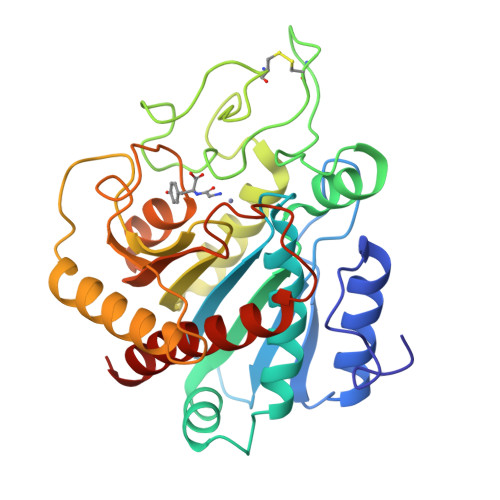
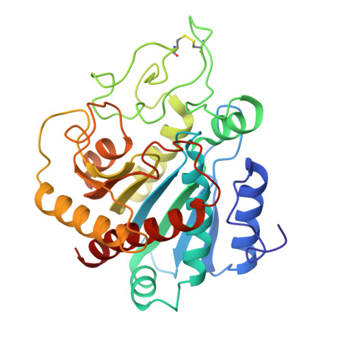
X-ray crystallographic investigation of substrate binding to carboxypeptidase A at subzero temperature.

Christianson, D.W.Lipscomb, W.N.

(1986) Proc Natl Acad Sci U S A 83: 7568-7572

  • DOI: https://doi.org/10.1073/pnas.83.20.7568
  • Primary Citation of Related Structures:  
    3CPA

  • PubMed Abstract: 

    A high-resolution x-ray crystallographic investigation of the complex between carboxypeptidase A (CPA; peptidyl-L-amino-acid hydrolase, EC 3.4.17.1) and the slowly hydrolyzed substrate glycyl-L-tyrosine was done at -9 degrees C. Although this enzyme-substrate complex has been the subject of earlier crystallographic investigation, a higher resolution electron-density map of the complex with greater occupancy of the substrate was desired. All crystal chemistry (i.e., crystal soaking and x-ray data collection) was performed on a diffractometer-mounted flow cell, in which the crystal was immobilized. The x-ray data to 1.6-A resolution have yielded a well-resolved structure in which the zinc ion of the active site is five-coordinate: three enzyme residues (glutamate-72, histidine-69, and histidine-196) and the carbonyl oxygen and amino terminus of glycyl-L-tyrosine complete the coordination polyhedron of the metal. These results confirm that this substrate may be bound in a nonproductive manner, because the hydrolytically important zinc-bound water has been displaced and excluded from the active site. It is likely that all dipeptide substrates of carboxypeptidase A that carry an unprotected amino terminus are poor substrates because of such favorable bidentate coordination to the metal ion of the active site.


Macromolecules
Find similar proteins by:  (by identity cutoff)  |  3D Structure
Entity ID: 1
MoleculeChains Sequence LengthOrganismDetailsImage
CARBOXYPEPTIDASE A307Bos taurusMutation(s): 0 
EC: 3.4.17.1
UniProt
Find proteins for P00730 (Bos taurus)
Explore P00730 
Go to UniProtKB:  P00730
Entity Groups  
Sequence Clusters30% Identity50% Identity70% Identity90% Identity95% Identity100% Identity
UniProt GroupP00730
Sequence Annotations
Expand
  • Reference Sequence
Small Molecules
Ligands 3 Unique
IDChains Name / Formula / InChI Key2D Diagram3D Interactions
TYR
Query on TYR

Download Ideal Coordinates CCD File 
C [auth A]TYROSINE
C9 H11 N O3
OUYCCCASQSFEME-QMMMGPOBSA-N
GLY
Query on GLY

Download Ideal Coordinates CCD File 
B [auth A]GLYCINE
C2 H5 N O2
DHMQDGOQFOQNFH-UHFFFAOYSA-N
ZN
Query on ZN

Download Ideal Coordinates CCD File 
D [auth A]ZINC ION
Zn
PTFCDOFLOPIGGS-UHFFFAOYSA-N
Experimental Data & Validation

Experimental Data

  • Method: X-RAY DIFFRACTION
  • Resolution: 2.00 Å
  • Space Group: P 1 21 1
Unit Cell:
Length ( Å )Angle ( ˚ )
a = 51.6α = 90
b = 60.27β = 97.27
c = 47.25γ = 90
Software Package:
Software NamePurpose
PROLSQrefinement

Structure Validation

View Full Validation Report


Entry History 

Deposition Data

  • Released Date: 1982-07-29 
  • Deposition Author(s): Lipscomb, W.N.
  • This entry supersedes: 1CPA

Revision History  (Full details and data files)

  • Version 1.0: 1982-07-29
    Type: Initial release
  • Version 1.1: 2008-03-25
    Changes: Version format compliance
  • Version 1.2: 2011-07-13
    Changes: Version format compliance
  • Version 1.3: 2017-11-29
    Changes: Derived calculations, Other