
|
|
|
|
|
||
PDB Community Focus: Julian Voss-Andreae, Protein Sculptor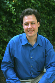
Photo by LeeAnn Gauthier. Julian Voss-Andreae is a German-born sculptor based in Portland, Oregon. In his youth he painted for a number of years, but then changed course and studied physics at the universities of Berlin and Edinburgh. After participating in a seminal quantum physics experiment in Vienna as part of his graduate research, Voss-Andreae moved to the US in 2000 with his passion for art rekindled. He graduated from the Pacific Northwest College of Art (PNCA) in 2004 with a BFA in sculpture. While still at PNCA, Voss-Andreae developed a novel kind of sculpture based on the structure of proteins, the building blocks of life. Voss-Andreae's work has been commissioned internationally and has been highlighted in journals such as Leonardo and Science.
Photographs of Voss-Andreae's sculptures, including those shown here, are now part of the RCSB PDB's Art of Science traveling exhibit. For more information on hosting this exhibit, please contact info@rcsb.org.
Your sculptures are amazing depictions of three-dimensional proteins. How do you decide upon the scale and the materials to use? Are you trying to tell a story about the molecule? Is there a connection between the proteins shown and the wood or metal? I want to make sculptures that work as metaphors. When I use the structure of a protein as a starting point, it is not only about the beauty of that structure. That can be accomplished equally well with a protein model. I want to create something meaningful beyond that. To give you an example, I created a sculpture based on the structure of collagen (using the coordinate file for 1BKV)1. I designed cutouts on each piece (a peptide unit) to reveal the dominant force lines, which is something we see in steel bridges and other steel constructions every day. That way the piece subtly alludes to its role in our body as a structural component. At some point I decided to depart from the molecular structure and opened up the intertwining helices toward the top. This expanded the meaning of the sculpture towards another important aspect most people think of in connection with collagen, aging. Collagen is responsible for our skin's elasticity – its degradation famously leads to the wrinkles that accompany aging. Together with the title "Unraveling Collagen" the piece now has the potential to function as a metaphor for our growth physically as well as mentally, and for our intertwined paths through life. 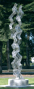 Julian Voss-Andreae
 Unraveling Collagen, 2005 Stainless steel, 24" x 135" x 32" In another piece, titled "Heart of Steel", I was interested in using a literal connection between the chemistry in the protein and the chemistry on the sculpture's surface. I made a complete human hemoglobin (1A3N)2 out of a certain kind of steel known as 'weathering steel'. This special alloy initially rusts like ordinary steel but eventually stops because the special oxide layer it builds up is not water soluble and thus protects it from further corrosion. I finished the piece with a shiny surface and installed it. Upon its unveiling it was still gleaming, but after a few rain showers the color started to change and within half a year it was dark red. What had completely changed the look of the sculpture is of course the same chemical reaction that occurs when we breathe: Iron binds to oxygen. 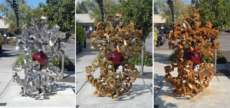 Julian Voss-Andreae
 Heart of Steel (Hemoglobin), 2005 Weathering steel and glass, height 5' Location: 1st Street/"A" Avenue, City of Lake Oswego, Oregon Another sculpture is based on a light-harvesting complex (1NKZ)3, the protein scaffolding containing a subunit of the photosynthesis apparatus in plants. So I displayed the piece on the floor of a dark empty room with a candle in the center casting shadows of the structures on the wall. That way the sculpture in its environment conveys a feeling of sacredness; it somehow resembles an altar. The flickering shadows of the eighteen alpha helices arranged in two concentric circles are intriguing, because they look a bit like moving plants. It is as if the light-harvesting complex still originates flora, but now with exchanged roles: The macroscopic plants of our world become ephemeral shadows, whereas the microscopic, and ordinarily not perceivable basis for their existence, becomes a tangible object. That is the kind of story I am interested in telling. It is about triggering associations and emotions. I want to make objects imbued with meaning of a poetic kind. My objects should have the potential to evoke metaphors in a scientific context which sometimes seems irreconcilable with the non-rational nature of poetry. 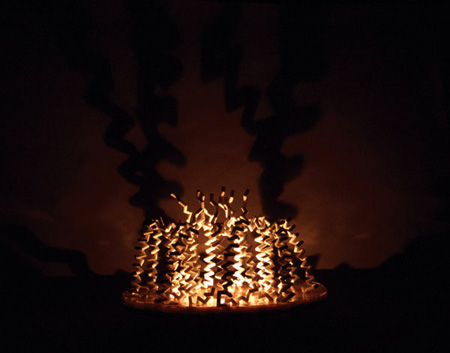 Julian Voss-Andreae
 Light-Harvesting Complex, 2003 Wood, casting resin, 22" x 25" x 25" How do you decide which proteins to sculpt? Is it the type of protein or the shape (helices, sheets, transitions of structural elements) that interests you? Sometimes I am fascinated mostly by the structure itself and sometimes more by the conceptual aspects, such as from what organism the protein originates and what biological role it plays there. A recent example for a sculpture based on an especially intriguing structure is a stainless piece portraying microcin J25 (1Q71)4. The small peptide resembles a lasso. Its tail folds over and goes through a noose made out of eight amino acids on the other end. An example of a protein mostly intriguing to me for its conceptual properties is hemoglobin. Ideally, both aspects are equally appealing. The first protein I saw, the green fluorescent protein (GFP), is such a molecule for me. It has a fascinating story as well as one of my favorite structures. It was actually an image of GFP that got me hooked on proteins in the first place while I was still doing science. I recently completed a stainless version that will possibly go on display at the Friday Harbor Laboratories on San Juan Island (Washington State), where GFP was isolated first in the early 1960's. I don't have a particular preference for certain types of proteins. Concerning secondary structure, I tend to like beta sheets better than alpha helices, because they interact more clearly with their neighbors whereas the helices look more isolated, which is illustrated by the position of the hydrogen bonds in those two motifs. The first large-scale outdoor sculpture I made was, however, a portrait of an alpha helix. This work, which was dedicated to the discoverer of the alpha helix, Linus Pauling, worked sculpturally very well though, because the helix was presented as an isolated object. I also like cyclic proteins very much. The first one I made was based on the plant cyclotide kalata B1 (1K48)5. It is a protein found in an African herbal medicine, with the effect of accelerating labor in childbirth. I made a large table top version of that protein that is now at the University of Queensland in Brisbane, Australia. Dr. David Craik has surrounded the sculpture with real Kalata-Kalata plants in pots, which I find a really beautiful idea. You wrote a computer program that takes a PDB coordinate file and indicates where the materials should be cut and rotated to build your sculptures. Was there a lot of trial and error in creating your early works? I wrote the core of that program in 2001 but I keep adding new parts. It allows me to make virtual renditions of the sculptures on the computer to troubleshoot before I build a real one; that worked very well from the start. The large challenge was never really the computer program but the translation into the real sculpture. In addition to inaccuracies there are also always the real, big mistakes we humans tend to make and it is surprising how many mistakes I made in my early work just because of the sheer number of measurements, cuts and connections. The latest addition to my program further eliminates sources of error by directly generating graphic files that can be used for computer-controlled cutting of metal with a laser beam or a thin water jet. Each peptide unit gets transformed into a four-paneled hinged piece of metal that comes complete with its number laser-etched on it. This approach significantly enhances the precision which allows me to tackle much larger proteins. The error still accumulates due to the one-dimensional nature of proteins, but it is small enough to be corrected on the way. Even though the laser-cutting replaces a lot of the manual labor and eliminates many errors, the construction is still an extremely time consuming process which requires a high level of skill. I still need about one hour of shop time per amino acid for a piece with amino acids the size of a hand. How close to the coordinate file are the final pieces? How important is scientific accuracy to your works? For pieces consisting of few amino acids, maybe up to 50, my sculptures are typically quite accurate in the sense that the center of each mitered-cut joint corresponds to the position of the carbon alpha atoms in the peptide chain. When the number exceeds about 100, corrections become necessary even though the cutting has an accuracy of a few percent and the connections are similarly precise. Heat distorts the metal and gravity kicks in at some point, resulting in unavoidable deviations from the exact molecular structure. Recently I started using much thicker material which holds up against gravity much better. But still, there are always some residual inaccuracies accumulating. I am not interested in accuracy just to be precise, but in order to capture the character of the protein structure. I don't mind if one end is off by a little bit with respect to the other, but it is unacceptable if some parts of the backbone have an unnatural spacing to their neighbors. If I have a beta sheet for example, I want to make sure that the zigzag pattern on neighboring strands corresponds to each other to capture the character of the fold. It is an important aspect of those pieces that these peptide chains don't just wiggle randomly in space but are in fact shaking around a delicately balanced configuration of minimal energy. In one case, when I made the sculpture based on kalata B1, I have asymmetrically stretched the coordinates a little bit. I needed some distortion in order to make my angles match up at the ends. But I did it in such a way that the orientation of my square tubing had the same Möbius strip topology as the backbone of the molecule. So it remained a faithful mapping of the topology, but not the coordinates. When I made a number of alpha helix-based pieces out of wood, I experimented with shortening the length of the peptide units proportional to the natural tapering of the wood, which yielded some very interesting results. For example, I built an alpha helix out of a tree cut into over 100 identical pieces decreasing in size. The condensation of the 30' (9 m) tree into the 10' (3 m) tall sculpture amplified the natural shape of the tree and caused the piece to have a striking resemblance to a human spine. I find such an unexpected emergence of a new level of meaning very interesting. My goal is to create a sculpture that works artistically. In some cases that might require an accurate mapping of the actual geometry, whereas in others it is only the specific departure from the exact geometry that allows it to become a meaningful piece of art. Which do you find more interesting – the shape of the protein, or the manipulation of these materials to make that shape? I love the structure of proteins and the diverse science behind it. I am stunned by the workings of this machinery of life. The aspect of my work I probably enjoy the most is that I get to explore and learn, and then dream up a sculpture, transforming my knowledge into something sensual and palpable, something that offers me, as well as others, an understanding on a deeper level. The most fulfilling is when a completed piece works well on many levels; esthetically, conceptually, and scientifically. And I really love getting feedback about my work from people I would have never met otherwise. How much information about science and technology do you include in your lectures to art students? In my lectures I talk about everything that fascinates me, and that is typically about 90% science. But I always try to present it as something sensual, with lots of visuals. I never really appeal purely to the intellect, but mostly to my audience's capacity to just watch and wonder. I do that not so much for pedagogic reasons, but because that is the way I function myself. Another aspect is that I always feel that there is a need to fill a gap of knowledge when it comes to science for non-scientists, especially in this country. I want to share my passion about science and show that it can be fun and extremely fascinating. We've noticed that people are interested in protein structures even when they don't know what they are looking at. However, that level of interest seems to greatly increase when they become aware of the specific protein being depicted. A picture becomes more engaging when it is known that it is, for example, hemoglobin. How does this play into your work? I have noticed that effect, too, especially with people who are generally interested in science. In fact, I used to display my work with labels talking a little bit about the science behind it. But I changed that because often the exact opposite happened, especially in the art world: As soon as you mention "science" people tend to shut off. They often feel uneasy with science and fail to recognize that it is a huge (and hugely important) part of human culture. And therefore, they rob themselves of the opportunity to see a whole world of beauty, the beauty revealed to us through scientific experiments. DNA is an iconic image at this point – it is recognized by almost everyone. Will structures of proteins follow the same path? I have my doubts. DNA is such a great image because of its simplicity. Take a ladder and twist it. But I do think that the general public is bound to get a clearer visual idea of protein structure in the future. Currently we are witnessing the merging of human technology and naturally evolved technology - biochemistry - and I find it likely that we will end up expanding on this existing technology in the long run instead of completely reinventing the wheel. So people will see many more proteins in the future I think. Right now people already see frequent images of proteins, mostly in cartoons illustrating for example lock-and-key interactions or in popular scientific illustrations of viruses. I think people often fail to understand at this point that all these images actually depict proteins. Maybe there are certain proteins that have the capacity for an iconic image; I could imagine, for example, the antibody. What are you working on now? I am currently working on three large commissions: Early next year I will be installing a 7' (2.10 m) stainless steel and glass version of a hemoglobin-based piece for a well-known scientist in Zurich, Switzerland. I have also started to work on a piece for Roderick MacKinnon in New York, who won the 2003 Chemistry Nobel Prize for elucidating the structure and function of the KcsA potassium channel protein (1K4C)6. He has commissioned me to create a sculpture inspired by that structure for his home. It will contain a blown glass object representing the internal space of the protein with its seven ion sites. And the new Scripps Research Institute in Florida has recently commissioned me to create a 12' tall stainless steel sculpture based on the antibody molecule which will be installed at the entrance of the new campus in Jupiter in 2008. That piece plays off of a very interesting formal correspondence I discovered between the proportions of the human body in Leonardo's famous "Vitruvian Man" from 1490 and the proportions of the antibody molecule as in the composite model by Eduardo Padlan.7 I have just finished fine-tuning the design and am in the process of having 1,500 pounds of stainless steel laser-cut. In addition to working with protein structure, I am very interested in finding sculptural metaphors for some aspects of quantum physics, which was my background before I turned back to art full-time. I recently finished the first version of a sculpture called "Quantum Man", a stylized image of a walking man consisting of about one hundred steel sheets oriented perpendicular to the direction of his motion. This piece was inspired by the question of how the wave function of a moving human would look like and it was covered by Science last August. Currently, I am working on a new version of that piece in bronze for a museum in Oregon. References:
|
||
©2007 RCSB PDB |