Education Corner
How Does Life Work? Creating Scientific Explanations in Stained Glass
By Joel Kowit, Ph.D.
Joel Kowit, Ph.D.
Distinguished Professor Emeritus of Biology at Emmanuel College, Boston, Joel Kowit is also Founder and Director of Immunology Workshops, and for three decades has trained research scientists in immunology at more than 40 pharmaceutical companies (www.immunologyworkshops.com) using several hundred scientific immunology illustrations which he has created. He has also been painting for 20 years and has had solo exhibitions at Cary Library, Lexington, MA; Emmanuel College, Gallery 5; The Massachusetts State House; and Lincoln Library, Lincoln, MA. Following a dozen years of sporadic glassblowing, Joel took up stained glass, and has applied his knowledge of science and his experience as an artist to a novel endeavor: Bio Stained Glass (www.biostainedglass.com), a synthesis of his experiences with graphic design, science, and art.
To use or copy any of these images, please request permission from the artist (kowitj@emmanuel.edu). Images may not be used for commercial purposes.
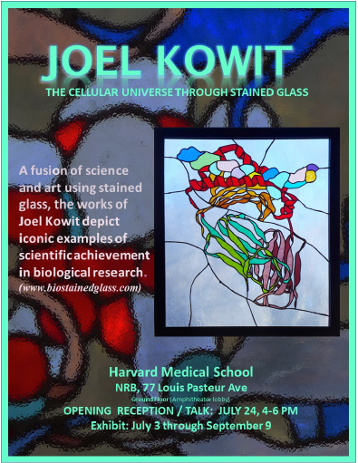
His exhibition The Cellular Universe Through Stained Glass will be on display July 3 through September 9 at the NRB/New Research Building at Harvard Medical School. An Opening Reception and Talk will take place July 24, 4-6 pm, 2019.
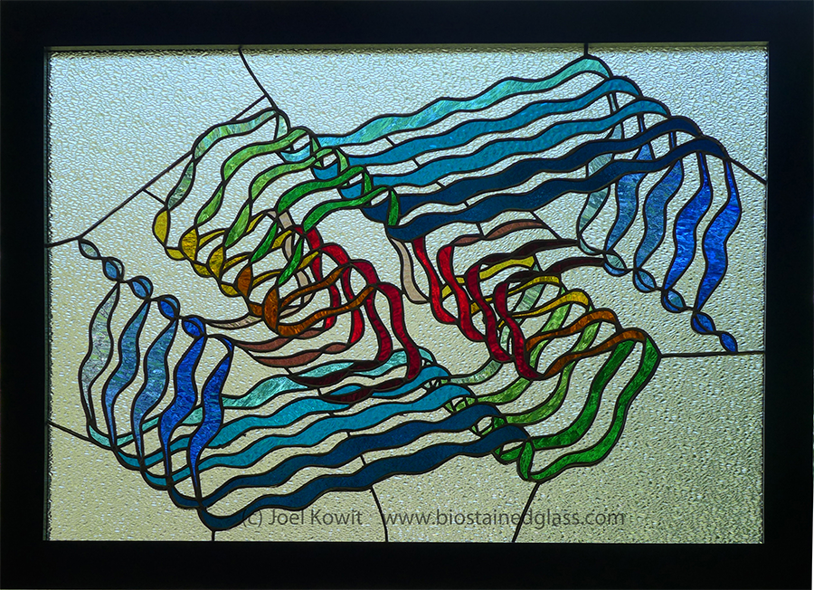
A stained-glass piece hangs in front of my dining room window – a strange conglomeration of over 70 cylinders, of various colors, with a snakelike ribbon twisting and winding its way through this maze of shapes. A visitor sees it for the first time: “It seems frenetic.” “Why those colors?” “It looks abstract.”

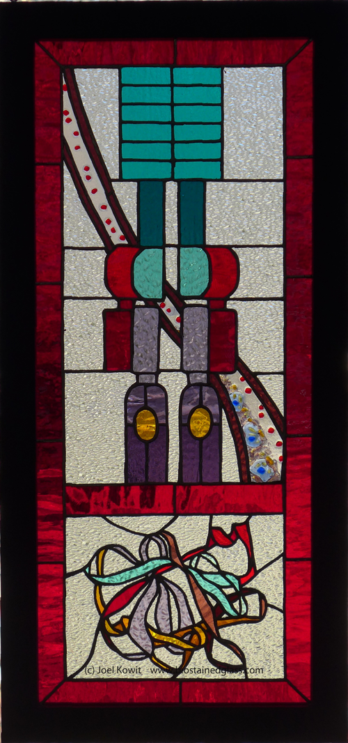
CRISPR Cas9 from PDB structure 4oo8 (H. Nishimasu et al. (2014) Cell 156: 935-949). Click to enlarge.
The piece, in fact, is a scientifically accurate representation of a protein, along with its bound, winding DNA and RNA, based on a Protein Data Bank entry for the protein (4oo8). This is a protein that has been in the news quite a bit lately–Cas9–the enzyme used in the process known as gene editing. With the sunlight coming through the glass, it sparkles and seems magical – a fitting metaphor for the amazing abilities of this enzyme to correct genes in living cells, and in living organisms.
Cas9 is one of eighteen stained glass pieces I’ve created in the three years since I retired as Professor of Biology, Emmanuel College, Boston. Though I had no formal training in art, I dabbled in drawing over the years, “sat in” on studio art courses in painting and in drawing at Emmanuel, and practiced glassblowing at MassArt and a local studio. I met a master stained-glass artist who incorporated his own blown works into his art. This led me to take a brief workshop in stained glass. The first step was choosing a subject. If one googles “stained glass images,” the works all seem to fall into only a few categories: religious, animals, portraits, flowers, landscapes, graphic designs. I chose to look elsewhere, and turned to my background for inspiration, and created a piece called Migration (Leukocyte Extravasation), based on the movement of white blood cells from blood into tissues at the site of infection–a subject I had lectured on in my immunology courses.
After completing this first stained-glass piece, I continued to take my inspiration from my own experiences as a scientist: something I worked on in the lab, taught in the classroom, or lectured about during my career. Several pieces are informed by work associated with Nobel Prizes in Chemistry and Physiology or Medicine; others are associated with important human diseases. All are scientifically accurate within the context of the art form, based on knowledge gained from years of study by researchers on the workings of these biomolecules and cellular structures.

Vibration (Organ of Corti) Sound transmitted into the inner ear causes vibration of the tectorial membrane (green). It impinges on a set of three outer and one inner rows of “hair cells.” They become activated and release neurotransmitters which activate neurons (blue) which transmit the information to the brain. Click to enlarge.
For example, Vibration (Organ of Corti) was inspired by my senior seminar, Medical Genetics. The course focused on three diseases: cystic fibrosis, Duchenne muscular dystrophy, and inherited deafness. A guest speaker was a deaf man who taught American Sign Language (ASL) to blind people, by touch! He spoke to the class through two interpreters about what it was like growing up deaf, and about the sophisticated grammar of ASL. It was a powerful and normalizing experience. Vibration is now in the collection of Decibel Therapeutics in Boston, a biotech company aimed at treating hearing disorders.
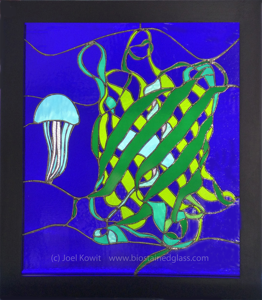
Excitation (Green Fluorescent Protein with Aequorea victoria). From PDB Structure 1gfl (F. Yang et al. (1996) Nat.Biotechnol. 14: 1246-1251). Click to enlarge.
Excitation (Green Fluorescent Protein with Aequorea victoria) was inspired by a sabbatical spent at the Transplantation Biology Research Center at Massachusetts General Hospital in the lab of a friend and colleague. One study involved following the fate of bone marrow cells transferred from green fluorescent protein (GFP) mice into “conditioned” non-GFP mice. (One of my own undergraduate students, an intern there, taught me how to obtain marrow from the tiny femur of a germ-free mouse.) The transplanted cells, which glowed green under a blue light, were characterized and studied by fluorescence-activated cell sorting. This is just one of many ways GFP, derived from a bioluminescent jellyfish, has been a powerful research tool in biology. (For their discovery, study, and application of GFP, Shimomura, Chalfie, and Tsien received the Nobel Prize in Chemistry in 2008.)
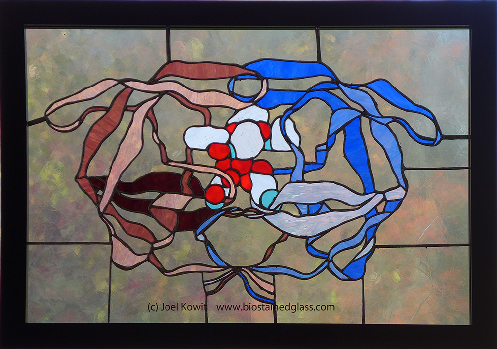
Inhibition HIV protease (ribbon model) with Merck’s inhibitor, Indinavir, in the active site. From PDB Structure 1hsg (Z. Chen et al. (1994) J.Biol.Chem. 269: 26344-26348).Click to enlarge.
The connection between a stained-glass piece and a research scientist can be very personal. One scientist who was at Merck in the 1980s and worked on the structure of the HIV protease and the development of Indinavir, wrote a comment on seeing the piece Inhibition:
The piece recalled a desperate time in human health history, and the dramatic response that inhibitor drugs like Indinavir brought about.
Inflammation (NLRP3 Inflammasome Series). Click to enlarge.
Sometimes, a piece is created in collaboration with a scientist. This was the case with the NLRP3 Inflammasome. A clinician, whose work focused on the association between atherosclerosis and inflammation, ran with his colleagues a clinical trial of over 10,000 patients, testing treatment with a monoclonal antibody to IL-1 beta (a cytokine with inflammatory effects.) The trial showed (among other things) that the treatment reduced atherosclerosis. He requested a stained-glass piece showing the inflammasome, the subcellular structure whose assembly is triggered by pro-inflammatory signals. Once assembled, it cleaves a precursor to produce active IL-1 beta. I designed a piece with an image showing the components of the inflammasome, a ribbon model of IL-1 beta in a lower panel, and in the background, a blood vessel with floating red blood cells and near the bottom, atherosclerotic plaque containing (bluish) white blood cells.
Other pieces have been shared with the research community. Degradation (26S Proteasome) appeared on the cover of Biochemical Journal in 2017. Tolerance (Major Histocompatibility Complex II with MBP peptide) was used to promote the 2019 EMBO workshop on Antigen Processing and Presentation.
Loss (Beta-amyloid fibril, Osaka mutation). From PDB structure 2mvx (A.K. Schutz et al. (2015) Angew.Chem.Int.Ed.Engl. 54: 331-335). Click to enlarge.
Scientific accuracy of the artwork is important. Colors are chosen, when possible, to relate to the science. For example, in Loss (Beta-Amyloid Fibril), the protein shown is a mutant form (Osaka mutation) associated with brain plaques from patients with early onset Alzheimer’s. I chose to show the colors of the 10 polypeptide units (five above, five below) fading as they go back in space in order to symbolize both the therapeutic approach of dissolving these fibrils as a possible treatment for Alzheimer’s, and the fading memories associated with this disease. “Loss” is in the collection of LabCentral in Cambridge, MA.
In Cas9, the cylinders, representing protein alpha-helices, are colored in groups corresponding to functional domains of the protein. The colors used for the bases are azure (adenine), turquoise (thymine), copper (cytosine), grape (guanine), and light turquoise (uracil.) I arranged the DNA sequence to encode cys-arg-ile-ser-pro-arg (C-R-I-S-P-R) and the unbound RNA sequence to encode an amino acid sequence (in single letters) which spells out all the initials of all the authors of the Nishimasu et al. Cell paper which solved the Cas9 structure 4oo8 (exceptions: glu for Z and Q for O).
Pieces showing cellular structures –Vibration (Organ of Corti), Vision (Retina), Degradation (26 S Proteasome), Contract (Muscle Sarcomere), Motion: 9+2 (Flagellar axoneme)–are designed based on our knowledge of the actual cellular structures and how they function.
When the subject of a stained-glass piece is a protein, the design begins with a PDB structure file. The protein structure is visualized using PyMol; I choose a view which is balanced, reflects my personal aesthetic, and minimizes potential problems with construction (such as many tiny pieces of glass). Corel Designer is used to recreate the design (by hand/mouse), sometimes coloring different segments to test out color schemes. A printout is made, enlarged to size, each individual segment labeled, and several copies printed. One copy is used as a layout on which the glass pieces will be assembled. A second is cut with a razor into every labeled segment. Each of these paper cutouts is traced onto glass (chosen for color and texture), and a hand glass cutter is used to cut the pieces. Loss (Beta-Amyloid Fibril) has over 300 pieces of glass; CRISPR Cas9, over 400. I keep a box of band-aids handy. The edges of the cut glass pieces are ground smooth, the pieces are copper foiled (with a thin adhesive backed copper tape), assembled, soldered on both sides, washed, and framed. Start to finish, a piece typically takes four months.
Finally, the artwork is hung in a window and viewed with natural light. With stained glass, it’s ALL about the light. And the light changes with the time of day, with the weather, the season, the skies, the position of the viewer, and whatever is in the background behind the glass ––snow, grass, a flowering tree. Sometimes the pieces glow and radiate color, sometimes they stay quietly subdued. This ever-changing light gives life to the works. This is a property of glass and no other medium. Additionally, the medium of stained glass parallels the microscope, which relies on the transparent surface of the cell to visualize light passing through stained internal structures.
I see all these works as testaments to science and to the researchers, their dedication, creativity, persistence; as a statement that says This is what science research is all about: the unraveling of a secret mechanism or puzzle, the satisfaction of discovery, sometimes the extraordinary reward of developing or contributing to a new therapy, to heal; to reveal the beauty of life. Each of the pieces has its own unique story, but all of my stained-glass pieces ask the same fundamental question: How Does Life Work?
The protein molecules and cell structures which are the subjects of these artworks are foreign to most non-scientists and evoke a sense of mystery and sometimes, wonder. This response invites engagement, not just to observe, but also to be curious, to question, to learn, to acquire some understanding, and hopefully appreciation, for the science behind the art. I want my artworks to make people curious enough to want to know more, and I want young people, who are the most curious, to start asking questions–especially questions that we never thought of and to which we don’t know the answers.