
|
|
|
|
|
||
COMMUNITY FOCUS: Shri C. Jain, RCSB Protein Data Bank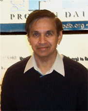
Shri C. Jain has been annotating structures for the PDB and the NDB since 1997. He has trained more than 25 members of the annotation staff and has annotated over one thousand structures. After receiving his PhD from the University of Poona, India, Dr. Jain taught at the Indian Institute of Technology Bombay, India for about two and half years and then continued his research at the University of Rochester, where he was a pioneer in the study of nucleic acid drug complexes. The focus of his X-ray crystallographic work was to study and understand how drugs, including intercalating compounds such as ethidium bromide, ellipticine, and acridine bind to nucleic acids. During that period, he solved the structure of the first actinomycin nucleoside complex [1]. This work was presented at the Cold Spring Harbor Symposium in 1971 -- the Protein Data Bank was founded at the same meeting. In 1992, he came to Rutgers The State University of New Jersey to join Dr. Berman's research group to continue structural studies on subtilisin and drug-nucleic acid complexes. In March 2006, he will retire after nearly 15 years of working at Rutgers. Q: You came to the PDB as a structural biologist. Tell us about your research. A: At Rochester, I solved structures of several drug-nucleic acid complexes in which the drugs such as ethidium bromide intercalate between the dinucleotide base pairs. The study of actinomycin-deoxyguanosine complex shed light on how it interacts with DNA. At Rutgers, I was involved with structural studies of the subtilisin-propeptide complex, actinomycin-nucleic acid complex, and alpha neurotoxin. Q: You've been annotating structures since 1997. Has it changed the way that you look at molecular structures? What types of challenges has annotation presented? A: When I first started annotating nucleic acid structures for the NDB in 1997, we were processing structures pretty much manually with the program MAXIT[2]. We did not have the data processing tools that we have now. A lot of automation has been incorporated since then to speed up the processing time with increased accuracy. The validation report that we send to depositors helps them evaluate their structure more closely so they can correct errors, if needed. We still visualize every structure with graphics programs such as RasMol [3, 4], (the internal program) Molly, or Chimera [5]. There has been a significant increase in the size and complexity of the structures that are being studied by the researchers and deposited with the PDB. Some of the very large structures, such as an icosohedral virus particle, have their own special beauty. Q: What has been the most challenging/most rewarding thing about annotation? A: Most of the time, annotation has been great fun. We get to see the latest trend in structural studies as these structures are deposited with PDB. But more importantly it gives a feeling of pride in being instrumental to providing an important service to the scientific community by processing structures accurately and consistently. It is not, however, as much fun when depositors do a poor job in deposition, or when an annotator is processing a huge complex structure in which a large part of the structure is defined as unknown, where several chains occupy alternate location, or the structure has a large number of new ligands. Q: How have the annotation tools changed over the years? A: Improvement in annotation tools is an ongoing process. There is a continuous demand for improved automation in data processing. The annotation tools available now have helped make processing structures easier and have minimized errors. Many diagnostic reports are created during the annotation process to continually warn us about what needs to be corrected. These tools and features, including the automatic creation of sequence database records, a ligand tool to process and add new ligands, and various scripts and MAXIT options have been of great help. Other tools, such the one that generates reminder letters to be sent to depositors, another that assists in updating structures on hold for publication, and all of the tools created to help prepare structures for public release have made the annotation process much easier. Q: Do you have any advice for future annotators? A: New annotators should pay close attention during training time and not be bashful in asking questions. Spend an hour or two everyday skimming over the data processing guide during training. Also review the PDB format guide to familiarize yourself with the record format. The new annotators need to be meticulous in record keeping. It will be very helpful to keep up with the status sheet that helps track down your workload. Don't be afraid to ask other members of the annotation staff for help or clarification. Enjoy processing structures – it's fun to use graphics programs to look at all of the different structures with their various shapes. Q: What will you miss about the PDB after retirement? A: Now that I am retiring, I will perhaps sometimes miss the daily grind of structure processing. Honestly, I think, what I will miss the most are all of the people that I have had an opportunity to work with for many years. It has been a great time and I am leaving with a sense of great pride that I had the opportunity to train so many annotators.
|
||
©2006 RCSB PDB |