DATA
QUERY, REPORTING AND ACCESS
Home Page Widgets
The entire RCSB PDB home page is comprised of customizable web widgets. Boxes with a dark blue bar on top are widgets that can be moved on the page by dragging the arrow buttons, hidden by selecting "Hide," or included in a customized view.
The  button in the top right corner lets users select which widgets are displayed by default. To only see query-related options, add widgets to download files and search by sequence and remove the Featured Molecules Widget that displays the RCSB PDB's Molecule of the Month and the Protein Structure Initiative's Featured Molecule.
Other widgets that can be displayed on the home page include the Comparison Tool for running pairwise structural and sequence alignments, links to ADIT for new and existing depositions sessions, and the New Features and Latest Structures widgets described below. button in the top right corner lets users select which widgets are displayed by default. To only see query-related options, add widgets to download files and search by sequence and remove the Featured Molecules Widget that displays the RCSB PDB's Molecule of the Month and the Protein Structure Initiative's Featured Molecule.
Other widgets that can be displayed on the home page include the Comparison Tool for running pairwise structural and sequence alignments, links to ADIT for new and existing depositions sessions, and the New Features and Latest Structures widgets described below.
New Website Features

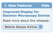
Descriptions of features added in any release can be accessed with
this widget.
Want to learn about the latest RCSB PDB tools and developments? The New Features Widget on the home page scrolls through the latest website applications and improvements. It also links to descriptions of all recent website releases.
Latest Structures
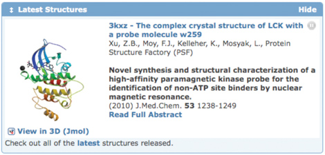
A new widget on the home page displays a slideshow of the latest PDB entries.
The Latest Structures Widget randomly cycles through all the entries that have been released in the most recent update. It displays the entry title, image, citation and PubMed abstract, if available. Users can pause the slideshow at any point to read the entire abstract, or click on the entry title to view the entry's Structure Summary page.

This widget rotates through all of the structures released in the most recent update.
Create a Collage of Structures
 After searching for structures, select Custom Report>Image Collage to display the set of returned structures as a series of tiled molecular pictures. Moving your mouse over each image in the collage displays the structure title; clicking on the small image shows a larger version. The PDB ID listed links to the corresponding Structure Summary page. After searching for structures, select Custom Report>Image Collage to display the set of returned structures as a series of tiled molecular pictures. Moving your mouse over each image in the collage displays the structure title; clicking on the small image shows a larger version. The PDB ID listed links to the corresponding Structure Summary page.
Image collages can be customized by the size of the images displayed and how many images are shown per page.
This image collage shows some of the structures that were returned for a search of structures that contained the same sequence as chain A of hemoglobin structure 4hhb.
Search the RCSB PDB in Your Web Browser

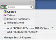
From Firefox, add RCSB PDB Search options by clicking on the arrow to the left of the search box.
In a hurry to search for a PDB entry? Add the RCSB PDB search engine to your web browser to perform queries directly through the browser's search box. RCSB Full Text or PDB ID Search and RCSB Author Search queries can be performed (alongside similar searches to Google and Yahoo) in the search box of modern browsers such as Firefox (versions 3+) and Internet Explorer (7+).
To add this OpenSearch functionality to your web browser:
• Visit www.pdb.org
• Click on the arrow near the search box
• For Firefox, select "Add RCSB Full Text or PDB ID Search/
Add RCSB Author Search"
• For IE, select the RCSB PDB search options from the "Add
Search Providers" menu
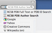
Then select one of the RCSB PDB search engines from the query pulldown menu and search the RCSB PDB directly from your browser.

Queries can be performed using this search box while browsing any website.
Reading about a PDB entry in a paper? Use the "RCSB Full Text or PDB ID Search" to search by the PDB ID. Use the "RCSB Author Search" to search for structures by the same author. As you type the author name in the browser search box, the "autocomplete" functionality will suggest possible name matches.
Turn Your Computer into a PDB Structure Kiosk
Highlight structures from your lab, institution, or class with the Molecules in Motion Kiosk Viewer. Using a list of PDB IDs, this full-screen animation program will display any PDB structure from different angles and perspectives. This Java viewer can be downloaded or launched from the RCSB PDB's Educational Resources page.

The kiosk program will also focus on and label any chemical components in an entry, like the heme group shown here in hemoglobin entry 4hhb.
Bookmark and Share RCSB PDB Webpages
 Easily send and store URLs by using the Share this Page button on the upper right side of all RCSB PDB webpages.
With this service, favorite PDB entries, Molecule of the Month features, and Looking at Structures pages can be emailed to colleagues or added to link sharing, bookmarking, and social networking sites such as Delicious, Facebook, and Twitter. Easily send and store URLs by using the Share this Page button on the upper right side of all RCSB PDB webpages.
With this service, favorite PDB entries, Molecule of the Month features, and Looking at Structures pages can be emailed to colleagues or added to link sharing, bookmarking, and social networking sites such as Delicious, Facebook, and Twitter.
Want to send an interesting PDB entry to a colleague? Use the Share this Page feature at the RCSB PDB site.
Website Statistics
Website access statistics for the second quarter of 2010 are given below.
Month |
Unique Visitors |
Visits |
Bandwidth |
April 2010 |
203139 |
486879 |
978.97 GB |
May 2010 |
199620 |
473973 |
1058.45 GB |
June 2010 |
174582 |
429274 |
869.39 GB |
|


