DATA
QUERY, REPORTING AND ACCESS
Website Statistics
Access statistics for the first quarter of 2011 are shown.
Month |
Unique Visitors |
Visits |
Bandwidth |
January |
210986 |
522651 |
982.26 GB |
February |
213862 |
526896 |
1114.34 GB |
March |
239640 |
598545 |
1004.99 GB |
Store Personal Annotations with MyPDB
 MyPDB lets users create a personalized version of the RCSB PDB accessible from any computer and from PDBMobile.
Using the MyPDB widget in the left-hand menu, users can create new accounts or log in. MyPDB lets users create a personalized version of the RCSB PDB accessible from any computer and from PDBMobile.
Using the MyPDB widget in the left-hand menu, users can create new accounts or log in.
- The MyPDB Saved Query Manager stores RCSB PDB searches (keyword, sequence, ligand, etc.) and composite queries built with Advanced Search. These saved queries can be run at the click of a button.
Stored searches can be set to run automatically. Email alerts (weekly or monthly) will be sent when matching entries are released.
- Personal Annotations. Users can save personal annotations and notes on the Structure Summary tab of any PDB entry, and add structures to a favorites list. The Personal Annotations summary page provides easy access to all of these tagged structures and annotations.
- User Account. Personal information (name, email address, account password, country, user type) can be updated at any time. All MyPDB account information is kept private and secure.
New Features at www.pdb.org
Improved Sequence tab features, new searches and reports, and a special educational view called PDB-101 have been added with the latest website release. See the What's New page for details and examples.
Structure Summary Pages
Every PDB entry has a Structure Summary page that offers a portal to tools, resources, and related links. Tabs on the initial summary page can be used to toggle between different topics. Users can also access different molecular viewers and external resources from these pages. A few of the many available options are highlighted below.
Quick Jmol Views for Exploring Macromolecular Structures

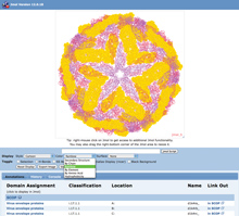
PDB entry 1k4r as seen in Jmol (jmol.sourceforge.net).The Jmol viewer is used by beginners and experts to interactively explore PDB structures.
Frequently-used Jmol options are available in pull-down menus and buttons, including the display style, color, and surface. For more detailed usage, click the right mouse button to access the Jmol menu, or enter scripts into the text box. Domain assignments from SCOP, DP, and PDP can also be displayed by selecting from the list provided. Select  from any entry's Structure Summary page to access the interactive viewer. from any entry's Structure Summary page to access the interactive viewer.
Structural Neighbors and the 3D Similarity Tab
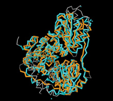
 Comparison of 1q6z (orange) with structural neighbor
3hww (cyan). Comparison of 1q6z (orange) with structural neighbor
3hww (cyan).
Proteins can have various degrees of similarity. If two proteins have highly similar amino acid sequences, it is generally assumed that they are closely related evolutionarily. As the evolutionary distance increases, the degree of similarity usually drops. Even if the sequence similarity is low, proteins may have similar functions and 3D structures. Detecting remote similarities, a core structural bioinformatics technique, is important in the study of functional and evolutionary relationships between protein families.
The RCSB PDB offers tools that quickly identify 3D protein sequence neighbors. For each PDB entry, the 3D Similarity tab lists the representative entries with 40% sequence similarity that are found using the jFATCAT-rigid1 algorithm. As an example, look at the 3D similarity tab for entry 1q6z. Representative protein chains are used since calculation of a real all vs. all comparison would require a great amount of CPU time.
Novel domain architectures and unexpected structural similarities can be detected by analyzing structural alignments. As an example, entry 3hww is one of the top ranking structural neighbors of 1q6z (chain A). Clicking on view from the Structure Similarity table will show a summary view of the alignment.
3hww has an RMSD of 3 Å based on the Cα positions, while the two protein chains are only 14% identical by sequence. 1q6z is a benzoylformate decarboxylase (EC number 4.1.1.7), while 3hww is a 2-succinyl-5-enolpyruvyl-6-hydroxy-3-cyclohexene-1-carboxylate synthase (EC number 2.2.1.9). Despite the low sequence identity and divergence in function, the high structural similarity indicates that both proteins evolved from a common ancestor.
Create Publication-Quality Molecular Images
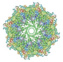
 Image of 3izi created using Protein Workshop Several interactive, Java-based2 tools can be used to visualize PDB data. Protein Workshop offers easily customized views; Simple Viewer offers a quick ribbon display; and Ligand Explorer visualizes the interactions of bound ligands in protein and nucleic acids structures. Image of 3izi created using Protein Workshop Several interactive, Java-based2 tools can be used to visualize PDB data. Protein Workshop offers easily customized views; Simple Viewer offers a quick ribbon display; and Ligand Explorer visualizes the interactions of bound ligands in protein and nucleic acids structures.
Each program can be used to create and save custom high-resolution images in JPEG, PNG, and TIFF formats. Using the Save Image dialog box from the File menu, users can specify the width and height of an image in pixels, inches, or millimeters.
Structural Biology Knowledgebase Widget
 The SBKB widget loads data about available models, targets, biological annotations, and more. Structure Summary pages host widget boxes of available data loaded from resources such as SCOP (scop.mrc-lmb.cam.ac.uk/scop) and CATH (www.cathdb.info). These boxes are in orange to indicate the data are annotations from external sources. The SBKB widget loads data about available models, targets, biological annotations, and more. Structure Summary pages host widget boxes of available data loaded from resources such as SCOP (scop.mrc-lmb.cam.ac.uk/scop) and CATH (www.cathdb.info). These boxes are in orange to indicate the data are annotations from external sources.
A new widget links to related information at the Structural Biology Knowledgebase (SBKB; sbkb.org), a free, comprehensive resource that integrates the results of structural biology and genomics efforts with other publicly available biological information to facilitate further research. The SBKB widget loads data about available models, protein targets, related biological annotations, related clones, and protocols with links to SBKB reports page.
References
- jFATCAT: Based on FATCAT (Y. Ye, A. Godzik (2003) Flexible structure alignment by chaining aligned fragment pairs allowing twists Bioinformatics 19: ii246-ii255), BioJava (R.C.G. Holland, T. Down, M. Pocock, A. Prlic, D. Huen, K. James, S. Foisy, A. Dr�ger, A. Yates, M. Heuer, M.J. Schreiber (2008) BioJava: an open-source framework for bioinformatics Bioinformatics 24:2096-2097), and Jmol (jmol.sourceforge.net).
- J.L. Moreland, A. Gramada, O.V. Buzko, Q. Zhang and P.E. Bourne (2005) The Molecular Biology Toolkit (MBT): A modular platform for developing molecular visualization applications. BMC Bioinformatics 6:21.
|


