|
|
|
|
|
|||||
PDB EDUCATION CORNER: USING VISUALIZATION TOOLS BY J. RICKY FOX, MURRAY STATE UNIVERSITY
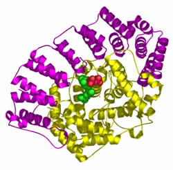
While a graduate student, I developed my love and passion for macromolecular structure, especially protein structure. It was also during this time that I fully realized the importance of noncovalent interactions in chemistry and biology. One of my favorite quotes while in graduate school was a paper from the lab of Professor Alan Fersht(1): "Biology is dominated by the chemistry of the noncovalent bond." This quote is on the wall of my office and my students would certainly agree that I emphasize noncovalent interactions in molecular systems. In the case of inter- and intramolecular interactions in biological molecules, it is very difficult for textbooks to present the diverse array of possible noncovalent interactions that can exist between functional groups and the role they play in the structure and function of macromolecular systems. This is why computer-based molecular visualization programs such as Protein Explorer, RasMol, Kinemage and Deep View have opened many doors for instructors and students to explore the structural nature of small and large systems. These programs allow the user to gain access to the wealth of information contained in the PDB. Although the structures in the PDB were not strictly obtained for educational purposes, chemistry and biology instructors can tap into this tremendous resource for pedagogical purposes. Currently, my students and I use Protein Explorer(2) to access the PDB, although one can visit the World Index of Biomolecular Visualization Resources (www.molvisindex.org) and choose a suitable program. Molecular Visualization in the Classroom I have been thoroughly impressed with how my colleagues have integrated the use of molecular visualization software and the PDB into the biochemistry and biology classroom.(3) Like many instructors that teach macromolecular structure, I utilize the PDB to teach the structural nature of these systems and to support material covered in the textbook. I also use Protein Explorer and the PDB to emphasize the nature and importance of noncovalent interactions that exist in proteins and protein complexes. Along with the normal classifications given to amino-acid side chains, I also categorize amino acids by their ability to form the various types of noncovalent interactions, especially the interactions involving aromatic systems (pi-pi interactions). pi-Type interactions in protein systems are not widely discussed in the biochemistry textbooks, even though they are an area of interest among the research community. Over the last several years, through my own mining of the PDB and student exploration projects, I have cataloged several examples of the different types of noncovalent interactions that exist between protein side chains and in protein-ligand complexes. I show these interactions during class when teaching protein structure and when I discuss the different types of complexes proteins can make with other biomolecules such as nucleic acids, carbohydrates and lipids. Even in the metabolism chapters, I try to show the structure of metabolic enzymes to support the discussion of the mechanistic features of these enzymes and the structure-function relationship that is so important in these chapters. In one biochemistry course I teach, the focus is on medicinal biochemistry, membrane structure and signal transduction. The students in this course have already completed a previous biochemistry course and are familiar with macromolecular structure. One might expect that the use of Protein Explorer and the PDB might be limited; however, this is certainly not the case. When teaching this course, I often find myself before lecture searching through the PDB looking for structures that can contribute to that day's lecture. I am usually successful at finding at least one structure in the PDB that can be shown and dissected via Protein Explorer that has direct application to the topic of the lecture. This type of approach leads to a balanced lecture period where there is an appropriate mix of lecture, student discussion and computer-based visualization. For example, a few days ago I was discussing the role of lipid tags (anchors) in the structure and function of peripheral membrane proteins. One method by which these proteins are associated with membranes is prenylation of C-terminal cysteine residues. This involves covalent modification with a farnesyl or geranylgeranyl group to produce a thioether linkage to the side chain of the cysteine residue. While looking through the PDB for a structure to discuss in class, I came across the PDB record (PDB ID: 1o1r) of a Protein Farnesyltransferase with bound geranylgeranyl diphosphate.(4) This alpha-helical dimer is a truly interesting and beautiful protein structure that caught my eye right away. I used Protein Explorer to visualize this structure in class as it was quite complementary to the discussion of lipid tags and it sparked a discussion on the structure and function of Ras proteins. I may have been able to show an image of this enzyme in class; however, the ability to manipulate this structure in Protein Explorer allowed me to show subtle aspects of the structure (three small beta-sheet regions) and to highlight some of the residues (Lys-164, Lys-294 and Arg-291 and Trp-303) involved in ligand binding. The basic residues interact with the phosphate groups of the geranylgeranyl diphosphate ligand via ionic interactions and the Trp side chain seems to be interacting with the hydrocarbon moiety of the ligand through CH-p interactions. The involvement of specific amino-acid residues in the structure and function of protein systems is always an important aspect of my biochemistry courses even though macromolecular structure might not be the major focus.
Student Use of Visualization and PDB Tools As a biochemistry instructor, I believe that utilizing computer-based visualization software, and data housed in the PDB, has been a tremendous asset in the teaching-learning process. I am very excited about the ability of students to use the software and the PDB data to explore macromolecular structure and search for noncovalent interactions. Over the past few years, I have had two different approaches to getting students involved in protein exploration projects. The first approach involves assigning individual students or student groups a particular macromolecule or giving them the option of finding their own structure in the PDB in which they have an interest. The students use visualization software to dissect the structure of the macromolecule and find several examples of noncovalent interactions important in stabilizing the structure of the protein or protein-ligand complex.5 The students present their findings to the class in oral presentations and field questions from me and other students in the class. Another approach I use, especially in larger classes, is to have students complete worksheets that contain questions and tasks concerning a particular structure from the PDB. These assignments are highly structured and require students to use Protein Explorer to investigate the structure of a particular protein or protein complex.3 The tasks and question for a particular assignment are provided below.
PDB Code: 1LOU These assignments are designed such that students have to acquire or recall information concerning the structures of amino acids and their ability to form noncovalent interactions. The students also have to demonstrate that they understand the distance and geometrical requirements for the formation of stabilizing interactions, which can be difficult when studying a macromolecule. The description required to complete the tasks and questions in the assignments involves measuring distances and manipulating the structures in Protein Explorer. Students quickly realize that proximity of amino-acid side chains does not guarantee that they are participating in a stabilizing (or repulsive) interaction. In my opinion, there is no way that traditional tests or quizzes can measure the ability of students to recognize and justify the existence of noncovalent interactions in protein systems. Other assignments I have used require students to comment on structural motifs that exist in proteins and to identify interactions involved in protein-ligand complexes. In some cases, students are asked to write a short paper on the biological function of protein system that is the focus of the assignment. In some of my biochemistry courses, I ask students to give oral presentations on the molecular basis of disease. To complement the information given on the various aspects of the diseases, students have searched the PDB to find relevant structures that can be visualized in Protein Explorer during the presentation. For example, a student recently gave a presentation on Phenylketonuria, a genetic disease linked to mutations in the enzyme phenylalanine hydroxylase (PDB Codes: 1PAH and 2PAH). Protein Explorer was used to show the multimeric nature of the enzyme, the catalytic and tetramerization domains, the catalytic iron ion and its coordinating side chains and the side chain of amino acids that are mutated in the disease-causing form of the enzyme. The use of molecular visualization in the student presentations was complementary to the other material presented and enabled the students to better explain the molecular basis of disease.
References: |
|||||
©2004 RCSB PDB |

