Education Corner
Creating sculptural models of proteins in a high-school engineering class
By Keagan O’Mara, Bioengineering Teacher, Bio-Med Science Academy STEM School
One of the hardest things about teaching biological engineering is getting students to understand and create mental models of biological concepts. So, for this project, my high school students had to build a physical model of a protein, complete with the proper primary structure. The goal was to give students a sense of how complicated proteins are, as well as to create models that could also function as sculptural art works and be aesthetically pleasing.
The original idea for this project came from a HASPI (Health and Science Pipeline Initiative, www.haspi.org) assignment. Students first started with learning about DNA and protein synthesis, and then moved into a series of videos which I sourced from YouTube explaining how the human body processes proteins from food and then turns amino acids into proteins. Students took notes in their Engineering Notebooks on the videos and sketched diagrams of the structure of amino acids, as well as the four levels of protein structure. Next, they moved into learning about the Protein Data Bank and how it is a powerful research tool. I created a very short overview hitting some of the key features of a protein summary page that students would need to know in order to create their projects.
For the purposes of this assignment, I limited students to a protein between 50 and 500 amino acids long. Students had to look at residue counts and determine what amino acid model they would create. Students were given a short list of suggestions as well as options to look through PDB-101 articles or search for themselves at RCSB.org.
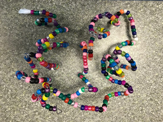
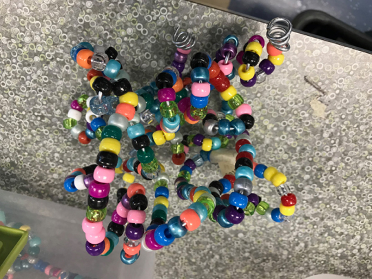
Once students had selected their proteins, they used the FASTA sequence for their entry to start to assemble the primary structure of their proteins. I used four 24-compartment organizer boxes with pony beads representing each of the standard amino acids, and a different color bead to represent each amino acid. Students strung the primary structure bead sequence on 14-gauge galvanized steel wire, then bent the wire into the secondary and tertiary structures as needed.
A useful tool which really came in handy was Autodesk’s MoleculeViewer.* This free (!) tool allowed students to zoom in on individual amino acid residues and view the structures in three dimensions! This tool was very handy in looking at amino acid sequences and seeing how individual residues related to one another in three-dimensional space.
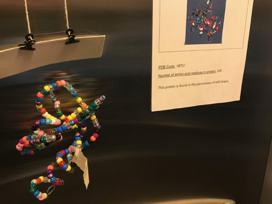
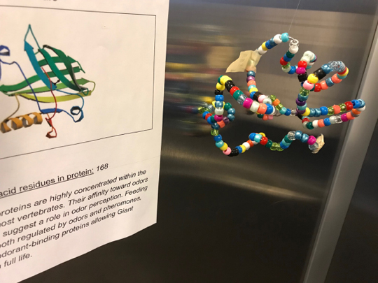
Finally, students created a display card that stated the name of the protein, had an image of the tertiary structure from RCSB PDB or MoleculeViewer, and also had information about the function of the protein. These cards were displayed with the completed models to help our audience understand what we were trying to accomplish.
The results from this project are a fascinating and beautiful display of protein shapes and forms that have garnered many compliments from my colleagues!
Here are some tips and hints from my experience with this project:
- Buy at least twice as many beads as you think you will need; it’s hard to gauge how many you will actually need, and some amino acids (like Lysine) will be used more often!
- I used 14 gauge galvanized steel wire because it seems to be in that sweet spot between easy to bend and shape, yet can handle a bit more abuse than pipe cleaners can.
- If I could go back in time, I would group my amino acid colors a little more purposefully (i.e. warm colors for hydrophobic, cool colors for hydrophilic, clear beads for essential amino acids); This might help students understand the connections between sequence and folding. You could also order colors to match the colors in MoleculeViewer!
- It isn’t possible to 100% replicate the forms of the protein secondary structure, but you can get pretty close. This is a great opening for a discussion on the drawbacks of model creation and the size/shape of amino acids! If you frame this assignment as an art project, you may find that hesitant science students will jump in full force.
* Learn more about the Autodesk Molecule Viewer from the Spring 2017 Education Corner by Merry Wang

Keagan O'Mara teaches Art and BioEngineering at Bio-Med Science Academy STEM school, a public STEM+Medicine high school on the campus of the Northeast Ohio Medical University. His work centers around integrating the visual arts into other subject areas and teaching BioEngineering concepts. He is interested in the intersection of science, engineering and the visual arts as well as using advanced technology in the classroom.
For more information and lesson materials, including supply lists and video links, please visit misteromara.weebly.com