Published quarterly by the Research Collaboratory
for Structural Bioinformatics Protein Data Bank

Fall 2009
Number 43

 
NEWSLETTER
  Contents  
Home | Newsletter Archive | PDF Version
Message from the RCSB PDB
Data Deposition
SF-Tool: A Tool for Crystallographic Experimental Data Validation
Meeting with Depositors at the ACA
Deposition Statistics
Structural Genomics News
Data Query, Reporting and Access
Improved Navigation of the RCSB PDB Website
Sequence Similarity Views of PDB Structures
New Tool For Exploring Sequence and Structure Alignments
Beta Release of Redesigned BioSync
wwPDB FTP Advisory Notice
Outreach and Education
Poster Prizes Awarded at ACA and ISMB
Recent and Upcoming Meetings and Presentations
Turn Your Computer into a PDB Structure Kiosk
Education Corner
Gary M. Battle, Ph.D.: Symposium on the Applications of Small-molecule Crystal Structure Information in Chemical Education
PDB Community Focus
Roland L. Dunbrack, Jr., Ph.D., Fox Chase Cancer Center
RCSB PDB PARTNERS, MANAGEMENT, AND STATEMENT OF SUPPORT


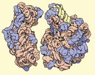
Message from the RCSB PDB

What's New?

Each week, approximately 140 structures and related experimental data are released into the PDB archive and loaded into RCSB PDB’s database.

To provide tools to query, report, and view these structures, the website at www.pdb.org is regularly updated with new features and resources. The button at the top of the each web page links to a description of the latest tools and enhancements. Detailed instructions, examples, and in some cases, screencasts illustrate how to access and use these features. Recent examples include new options available for reviewing query results and the Comparison Tool for calculating pairwise sequence and structure alignments.

Detailed news about all RCSB PDB activities is published online each week, and in this quarterly newsletter.

We welcome your questions and comments about these new features.

Nobel Prize for the Ribosome


Three structural biologists have won the 2009 Nobel Prize in Chemistry for studies of the structure and function of the ribosome–Venkatraman Ramakrishnan (MRC Laboratory of Molecular Biology), Thomas A. Steitz (Yale University), and Ada E. Yonath (Weizmann Institute of Science). The depositions of their first complete ribosome subunit structures (PDB IDs 1fjg, 1ffk, and 1fka) almost a decade ago ushered structural biology into a new era.

Since that time, more than 120 ribosome structures consisting of 50S, 30S subunits and complete 70S ribosomes have been contributed by these Nobel scientists. The structures, complexed with and without antibiotics, tRNAs, mRNAs, initiation factors, and release factors, provide a basis for understanding how the ribosome works and are useful tools for drug development.

Ribosome-related resources at the RCSB PDB include the October 2000 edition of the Molecule of the Month, animated GIFs of the large and small ribosomal subunit, and a small poster commemorating the award.

 
Snapshot: October 1, 2009
60464 released atomic coordinate entries
Molecule Type
55917
proteins, peptides,
and viruses
2060
nucleic acids
2454
protein/nucleic acid
complexes
33
other
Experimental Technique
52017 X-ray
8043
NMR
254
electron microscopy
18
hybrid
132
other
 
41210
structure factor files
5329
NMR restraint files
  Participating RCSB Members: Rutgers • SDSC/SKAGGS/UCSD
E-mail: info@rcsb.org • Web: www.pdb.org • FTP: ftp.wwpdb.org
The RCSB PDB is a member of the wwPDB (www.wwpdb.org)
©2009 RCSB PDB