DATA
QUERY, REPORTING AND ACCESS
Improved Navigation of the RCSB PDB Website
www.pdb.org has been reorganized to make navigating the website and search results easier and more intuitive.
The left-hand menu now groups frequently-used web pages into sections–Deposition, Home, Search, Tools, and Education–that can be moved up and down to create a left-hand menu ordered by user interest. This customized menu will then appear on every webpage. Links that appeared in the old left-hand menu system have been moved to more contextual places, such as query result pages.
By default, search results now display the most recently-released
structure first. Results can also be sorted by PDB ID, Residue Count, and Resolution.
Query result pages offer different tabs for reviewing any Structure Hits, Unreleased Structures, Citations, Ligand Hits, Web Page Hits, and GO, SCOP, and CATH Hits. Options for viewing, downloading, and generating reports for these search results appear as icons at the top of each page. Mousing over the icon indicates what action will be performed. A detailed description of the new result browser options is available from the What’s New page at www.pdb.org.
Sequence Similarity Views of PDB Structures
Interested in finding homologous protein structures, or finding a non-redundant set of proteins? The new Sequence Similarity View is an easy way to get this information based upon a protein chain in a PDB entry. This view, available through a tab at the top of each Structure Summary page, provides clusters of structures at different levels of sequence similarity. Details about the structures of all homologous proteins found within a cluster can then be examined.
The clusters are based on a weekly BLAST analysis of all proteins with more than 20 amino acids in the PDB. Many PDB entries contain several chains, so the sequence similarity is defined on a chain-by-chain basis, with the results returned for the entire structure. For more information, see the description at www.pdb.org/pdb/statistics/clusterStatistics.do.
New Tool For Exploring Sequence and Structure Alignments


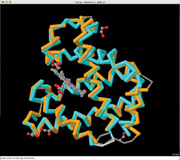
FATCAT structure alignment for 1mgn and 2nrl viewed using the JFatCat tool
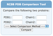
The new RCSB PDB Comparison Tool can calculate pairwise sequence and structure alignments using a variety of methods. This feature is available on the Compare Structures web page and as a downloadable web widget.
This functionality is also integrated with the Sequence Clusters offered from each entry's Sequence Similarity tab. Users can select a pair of chains from a given sequence cluster, and then run either sequence or structure alignments.
The current sequence alignments possible are: blast2seq,1 Needleman-Wunsch,2 and Smith-Waterman;3 the structure alignments offered are FATCAT,4 Mammoth,5 TM-Align,6 and TopMatch.7 FATCAT alignments can be viewed on the external server at fatcat.burnham.org/
fatcat or through the RCSB PDB's new JFatCat tool. JFatCat is a 3D structure alignment program based on FATCAT, BioJava8 and Jmol.9 JFatCat can be launched from the Comparison Tool or downloaded to the desktop.
Beta Release of Redesigned BioSync
 Synchrotron beamlines account for close to 80% of current X-ray
structures deposited to the PDB. The new capabilities and descriptions
of operational synchrotron beamlines worldwide are now available at
biosync-beta.rutgers.edu. The new site also offers search functions for services and equipment. BioSync has been redesigned to include the dramatic changes in data collection capabilities at synchrotron beamlines (including remote data collection, mail-in, crystallization and structure solution services, robotics handling for crystal screening and mounting, microfocus beams and facilities for collecting data under extreme conditions). The new look and feel of the website helps users find information about particular beamlines and to search for capabilities, services and equipment. The website's enhanced interface lets synchrotron personnel dynamically edit and add data. Synchrotron beamlines account for close to 80% of current X-ray
structures deposited to the PDB. The new capabilities and descriptions
of operational synchrotron beamlines worldwide are now available at
biosync-beta.rutgers.edu. The new site also offers search functions for services and equipment. BioSync has been redesigned to include the dramatic changes in data collection capabilities at synchrotron beamlines (including remote data collection, mail-in, crystallization and structure solution services, robotics handling for crystal screening and mounting, microfocus beams and facilities for collecting data under extreme conditions). The new look and feel of the website helps users find information about particular beamlines and to search for capabilities, services and equipment. The website's enhanced interface lets synchrotron personnel dynamically edit and add data.
BioSync offers deposition statistics by each synchrotron site and by geographical region. Galleries of structures and tables containing citations and other general information (e.g., phasing methods, resolution, R-factors, numbers of atoms) are also available. A separate set of statistical tables, galleries and informational tables is provided for structures produced by structural genomics efforts.
The redesign and upgrade of BioSync is being funded by NIGMS.
wwPDB FTP Advisory Notice
Four changes will be made to the wwPDB FTP site on November 24, 2009.
- The script for mirroring the FTP site using the rsync program will
be modified to prompt the user to choose one of three rsync servers
(RCSB PDB, PDBe, PDBj).
- The top-level README file will point to the download instructions
hosted on the wwPDB website
- The directory of newsletters will be updated
- Sequence cluster data that is used only by the RCSB PDB website
will be removed from the wwPDB FTP site. The data will be made available from the RCSB PDB at ftp://resources.rcsb.org/
sequence/clusters/.
Detailed information can be found at www.wwpdb.org.
1. T.A. Tatusova & T.L. Madden. (1999) BLAST 2 Sequences, a new tool for comparing protein and nucleotide sequences. FEMS Microbiol Lett 174:247-250.
2. S.B. Needleman & C.D. Wunsch. (1970) A general method applicable to the search for
similarities in the amino acid sequence of two proteins. J Mol Biol 48:443-453.
3. T. F. Smith & M. S. Waterman. (1981) Identification of common molecular subsequences. J Mol Biol 147:195-197.
4. Y. Ye & A. Godzik. (2003) Flexible structure alignment by chaining aligned fragment pairs
allowing twists. Bioinformatics 19:ii246-255.
5. A.R. Ortiz, C.E. Strauss & O. Olmea. (2002) MAMMOTH (matching molecular models
obtained from theory): an automated method for model comparison. Protein Sci 11:2606-2621.
6. Y. Zhang & J. Skolnick. (2005) TM-align: a protein structure alignment algorithm based on the
TM-score. Nucleic Acids Res 33:2302-2309.
7. M. J. Sippl & M. Wiederstein. (2008) A note on difficult structure alignment problems. Bioinformatics 24:426-427.
8. R.C. Holland, T.A. Down, M. Pocock, A. Prlic, D. Huen, K. James, S. Foisy, A. Drager,
A. Yates, M. Heuer & M.J. Schreiber. (2008) BioJava: an open-source framework for
bioinformatics. Bioinformatics 24:2096-2097.
9. Jmol: an open-source Java viewer for chemical structures in 3D. http://www.jmol.org/
|


 Synchrotron beamlines account for close to 80% of current X-ray
structures deposited to the PDB. The new capabilities and descriptions
of operational synchrotron beamlines worldwide are now available at