for Structural Bioinformatics Protein Data Bank
Summer 2009
Number 42
DATA QUERY, REPORTING AND ACCESS Website access statistics for the second quarter of 2009 are given below.
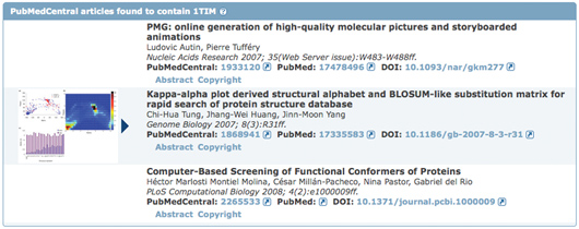
Literature View: Looking at Structures in PubMedCentral The recently-released Literature View aims to provide a broad look at how a given structure has been analyzed and presented in open access journals. For these publications, the full text of the article is available without copyright restriction. The overall intent of this feature is to increase awareness of all publications associated with the structure under study. Accessible by selecting the Literature tab from any Structure Summary page, this view highlights a variety of published articles associated with a PDB entry. The primary citation's abstract is included and can be used to search for structures with PubMed abstracts containing the same keywords. The Literature View also indicates the other publications included in the entry by the authors in addition to the primary citation (those contained in REMARK 1). In collaboration with the BioLit project (biolit.ucsd.edu), the Literature View lists any open access articles found in PubMedCentral that contain the entry's PDB IDeven the papers that do not include the PDB ID's primary citation in the references. Links to the abstract and copyright information, along with figures and related legends from these articles are displayed. If the listed open access articles also mention other PDB entries, their image, PDB ID, title, and sequence similarity to the initial structure are included in a table with links to their Structure Summary pages.
Customizable Structure Summary Pages Tabbed Structure Summary pages let users explore information about any given structure. Located above the structure ID and title, each tab offers a different type of informationa general overview, derived data, sequence details, sequence similarity, literature view, biology & chemistry, methods, geometry, and links to external resources. Data are organized on each page in different blocks, or "widgets". On a structure's Summary and Annotations pages, these widgets can be hidden or expanded. The positioning of selected widgets can be moved around on the page. This feature lets users organize these pages to highlight information specific to their interests.
An online video demonstrating how to customize these page layouts is available from the RCSB PDB site in the Whats New section. Customized layouts are automatically stored in a cookie and are retained for all PDB IDs. To switch back to the default layout, users should click on the "Reset View" in the left-hand menu. These tabs organize all of the different types of information available about each structure into focused web pages.
These tabs organize all of the different types of information available about each structure into focused web pages. MyPDB: Keep up-to-date with new
structures...automatically!
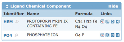
Ligand Expo: Searching and Browsing Features Ligand Expo (ligand-expo.rcsb.org) can be used to access chemical and structural information about all small molecule components found in PDB entries. The latest release includes amino acid variantsprotonation and de-protonation statesas part of Ligand Expo's Browse option. For example, when browsing arginine, protonation states ARG_LEO2 and ARG_LFOH_RNH1 will be included in the results list. Ligand Expo can also be used to search for ligands using:
Ligand Expo is based upon the Chemical Component Dictionary (www.wwpdb.org/ccd.html) maintained by the wwPDB. Ligand Expo's capabilities have been described in a flyer available for download from www.pdb.org. |
E-mail: info@rcsb.org • Web: www.pdb.org • FTP: ftp.wwpdb.org
The RCSB PDB is a member of the wwPDB (www.wwpdb.org)