Outreach and Education 
Celebrate DNA Day on April 25
Commemorate the 20th anniversary of the Human Genome Project's completion and the 70th anniversary of the discovery of the DNA double helix with PDB-101 resources
- Creating a paper model of DNA (available in English and Spanish from PDB-101)
- Visiting the National Human Genome Research Institute's DNA Day resources
- Extracting DNA in your kitchen (strawberries recommended; you can pound them in a plastic bag instead of using the blender)
- Exploring Molecule of the Month features on DNA, Designed DNA Crystal, DNA Polymerase, and more at PDB-101

Download images of DNA and other molecules to add a backdrop to your next virtual meeting.

DNA and RNA are the cell’s way of storing and deploying genetic information. Structural biology is revealing that some nucleic acids also fold to form complex molecular machines. Guided by these structures, nanotech scientists are building new machines composed of nucleic acid. Learn more at PDB-101.
New Coloring Page: Myoglobin in a muscle cell
The Biologist magazine hosts the Big Biochemical Colouring-in Series to invite readers to get artistic while learning about some of the life sciences’ most important and interesting macromolecules.
The latest feature on Myoglobin in a Muscle Cell has been published and made available at PDB-101.
This image shows the inner structure of a whale muscle cell. This cross section shows the space between two muscle sarcomeres, which are shown at the top and bottom, with actin and myosin filaments, which together provide the force for moving muscles. Glycogen granules, at the upper left, store glucose. The center portion is filled with glycolytic enzymes that generate usable energy from glucose and many small myoglobin molecules that store oxygen during the whale’s deep dives. A tubule of the sarcoplasmic reticulum that helps coordinate the timing of muscle contraction is shown in cross-section at lower right.
Many thanks to the Royal Society of Biology for sharing these pages with PDB-101.
PDB-101 hosts a collection of molecular-themed coloring books.
Paper Published: EM Holdings of the PDB

3DEM structures (PDB and EMDB) and density maps (EMDB only) versus time.
RCSB PDB published a review of 3DEM holdings in a special issue of Biophysical Reviews dedicated to Haruki Nakamura. Prof. Nakamura was one of the founders of the Worldwide Protein Data Bank.
Electron microscopy holdings of the Protein Data Bank: the impact of the resolution revolution, new validation tools, and implications for the future (2022) Biophys Rev 14: 1281–1301 doi:10.1007/s12551-022-01013-w
Molecular Landscapes

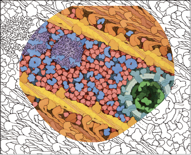
The aquatic bacterium Caulobacter crescentus has a two-step life cycle: it is born as a free-swimming “swarmer” and differentiates into an immobile “stalked” cell. As part of this process, it populates the two ends of the cell (poles) with macromolecular complexes that regulate these two different life forms. These specialized polar complexes in turn guide construction of flagellar motors and pili in swarmers and a holdfast structure in stalked forms, and regulate replication and segregation of the DNA chromosome as the cell grows and divides.
A distinctive microdomain, formed by the disordered protein PopZ, has the job of marking the poles and gathering these macromolecular complexes (“client” proteins) that have pole-specific functions. PopZ forms a condensate (yellow) that acts as a defined membraneless microdomain, excluding large cytoplasmic molecules like ribosomes from the pole, and selectively recruiting client proteins.
In this illustration, the microdomain is interacting with several soluble clients involved in regulation (orange), several membrane-bound clients (yellow-green), and DNA binding proteins (magenta).
This painting is part of PDB-101's SciArt gallery of Molecular Landscapes by David S. Goodsell.
Congratulations to Shuchismita Dutta
Dr. Shuchismita Dutta has been named a Fellow of the American Association for the Advancement of Science.
Visit the AAAS to learn more about the 2022 Fellows.

Dr. Shuchismita Dutta. Photo: Nick Romanenko/Rutgers University.